
Page 635 - Equine Clinical Medicine, Surgery and Reproduction, 2nd Edition
P. 635
610 CHAPTER 3
VetBooks.ir 3.30 3.31
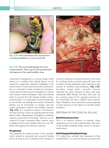
Fig. 3.30 Oral examination with a dental mirror
revealing infundibular necrosis of tooth 209.
Fig. 3.31 Post-mortem photograph of a severe
oroantral fistula. There is gross food contamination
and empyema of the caudal maxillary sinus.
conservative management or, at most, lavage of the recurrence indicates continued infection of the sinus
sinuses via a trephine hole. Dental disease can be by a leaking fistula, persistent diseased tissue such
treated by conservative dentistry techniques or by as dental or bone fragments and/or infection in the
tooth removal. Tooth removal is by either oral extrac- alveolus or maxillary bone/turbinates (Fig. 3.32).
tion or minimally invasive transbuccal techniques. Secondary fungal and/or anaerobic bacterial
Tooth removal under general anaesthesia is unusual infections are quite common in these cases post-
with modern dental techniques. Once the infected operatively. Most fistulae will heal with time, but
tooth is removed then the sinusitis usually resolves, the economic aspect of the ongoing treatment can
provided there is no subsequent oroantral fistula due be considerable and lead to difficult client relation-
to the alveolus not healing post removal. Oroantral ships. Neoplastic sinus disease has a poor prognosis
fistulae can be frustrating to manage and may as most tumours in this region are invasive and/or
require prolonged treatment and multiple surgeries malignant.
(Fig. 3.31). It is essential to achieve a seal between
the sinus and oral cavities by packing material in the EMPYEMA OF THE CONCHAL BULLAE
dental socket. Management of neoplastic secondary
sinusitis is generally unrewarding. Tumours can be Definition/overview
removed by large frontonasal flaps; however, most This is an unusual variation of sinusitis, where
are highly invasive and complete removal is very dif- inspissation occurs in the conchal bullae, the centre
ficult, with aggressive recurrence common. of the scrolled turbinate bones, rostral to the para-
nasal sinuses.
Prognosis
The prognosis for dental sinusitis is fair, provided Aetiology/pathophysiology
tooth removal is successful and oroantral fistulae CT studies have revealed that inspissation of the
do not develop. Lack of resolution of the sinusitis or conchal bullae is a common finding associated with