
Page 630 - Equine Clinical Medicine, Surgery and Reproduction, 2nd Edition
P. 630
Respir atory system: 3.2 Surgical conditions of the respir atory tr act 605
VetBooks.ir Differential diagnosis 3.19
The most important differential diagnosis is second-
ary sinusitis, particularly that due to dental disease.
Other causes of unilateral nasal discharge include
guttural pouch disease, particularly empyema, rhi-
nitis (particularly fungal rhinitis) and occasional
horses with mucopurulent tracheal discharges that
are preferentially expelled down one nostril.
Diagnosis
Diagnosis is based on the history, clinical signs, 6
endoscopy, sinuscopy and radiography. Resonance
on percussion over the sinuses may be reduced but 5
is an unreliable test. The mouth requires careful 4
examination for any cheek tooth pathology, most 3
particularly in the three caudal upper cheek teeth.
Endoscopy is less valuable in sinusitis than in many 2
other conditions of the URT, but it should be pos- 1
sible to establish whether there is any conchal swell-
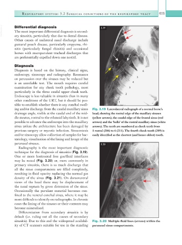
ing and/or discharge from the caudal maxillary sinus Fig. 3.19 Laterolateral radiograph of a normal horse’s
drainage angle, visible at the caudal end of the mid- head, showing the rostral edge of the maxillary sinuses
dle meatus, rostral to the ethmoid labyrinth. It is not (yellow arrows), the caudal edge of the frontal sinus (red
possible to advance the endoscope into the maxillary arrows) and the ‘bulla’ of the rostral maxillary sinus (white
sinus unless the architecture has been damaged by arrows). The teeth are numbered as cheek teeth from
previous surgery or mycotic infection. Sinocentesis 1 rostral (206) to 6 (211). The fourth cheek tooth (209) is
and/or sinoscopy allow collection of samples for bac- easily identified as the shortest (and hence oldest) tooth.
teriology, visualisation of the lining and lavage of the
paranasal sinuses. 3.20
Radiography is the most important diagnostic
technique for the diagnosis of sinusitis (Fig. 3.19).
One or more horizontal free gas/fluid interfaces
may be noted (Fig. 3.20) or, more commonly in
primary sinusitis, there is so much discharge that
all the sinus compartments are filled completely,
resulting in fluid opacity replacing the normal gas
density of the sinus (Fig. 3.21). On dorsoventral
views of the head there may be displacement of
the nasal septum by gross distension of the sinus.
Occasionally the purulent material becomes con-
fined to the ventral conchal sinus, where it may be
more difficult to identify on radiographs. In chronic
cases the lining of the sinuses or their contents may
become mineralised.
Differentiation from secondary sinusitis is by
default (i.e. ruling out all the causes of secondary
sinusitis). Due to this and the widespread availabil- Fig. 3.20 Multiple fluid lines (arrows) within the
ity of CT scanners suitable for use in the standing paranasal sinus compartments.