
Page 627 - Equine Clinical Medicine, Surgery and Reproduction, 2nd Edition
P. 627
602 CHAPTER 3
VetBooks.ir has been reported. An alternative approach to the These are mainly secondary invaders of tissue dam-
aged by trauma, sinonasal surgery, nasal or sinus
caudal aspect of the atresia is via a laryngotomy.
Prognosis masses such as progressive ethmoidal haematoma,
or persistent nasal discharge, either from disease
Prognosis for survival of unilateral cases is good of the paranasal sinuses or the lungs. Primary
but for return to normal exercise tolerance is poor. fungal rhinitis is very rare, particularly in temperate
Concurrent congenital abnormalities may seriously climates.
affect the long-term prognosis.
Clinical presentation
FUNGAL RHINITIS Fungal rhinitis results in a chronic, unilateral, nasal
discharge, often malodourous with variable amounts
Definition/overview of blood and a mucopurulent or purulent character.
Fungal rhinitis is a frequent complication of other The discharge may be ignored by the client until
nasal or sinus disease, or their treatment. The it becomes bloodstained. If the disease follows sur-
condition is predisposed (as with most fungal infec- gery, there may be a worsening or persistence of the
tions) by treatment with antibiotics or antiseptics. discharge. There may be submandibular lymph node
The other feature of fungal infections, the erosive enlargement.
nature of the fungal plaque, is also pertinent to this
disease. Primary fungal infections of the URT that Differential diagnosis
are caused by specific fungal species are rare and Other causes of chronic nasal discharge and epi-
tend to occur in certain parts of the world. Such staxis including progressive ethmoidal haematoma,
infections include cryptococcosis, rhinosporidiosis, guttural pouch disease and other paranasal sinus
phycomycosis and coccidioidomycosis. diseases should be considered. Fungal rhinitis is a
complication, rather than a differential diagnosis, of
Aetiology/pathophysiology most causes of nasal discharge.
The most commonly isolated fungus is Aspergillus
fumigatus but Pseudoallescheria boydii has been cultured. Diagnosis
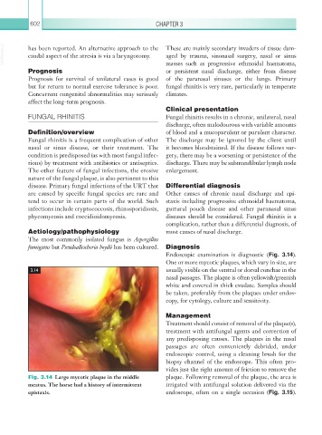
Endoscopic examination is diagnostic (Fig. 3.14).
One or more mycotic plaques, which vary in size, are
3.14 usually visible on the ventral or dorsal conchae in the
nasal passages. The plaque is often yellowish/ greenish
white and covered in thick exudate. Samples should
be taken, preferably from the plaques under endos-
copy, for cytology, culture and sensitivity.
Management
Treatment should consist of removal of the plaque(s),
treatment with antifungal agents and correction of
any predisposing causes. The plaques in the nasal
passages are often conveniently debrided, under
endoscopic control, using a cleaning brush for the
biopsy channel of the endoscope. This often pro-
vides just the right amount of friction to remove the
Fig. 3.14 Large mycotic plaque in the middle plaque. Following removal of the plaque, the area is
meatus. The horse had a history of intermittent irrigated with antifungal solution delivered via the
epistaxis. endoscope, often on a single occasion (Fig. 3.15).