
Page 629 - Equine Clinical Medicine, Surgery and Reproduction, 2nd Edition
P. 629
604 CHAPTER 3
VetBooks.ir 3.16a 3.16b 3.16c
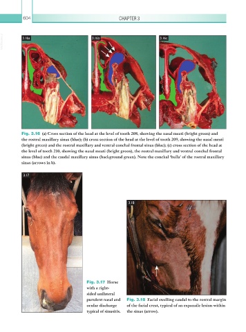
Fig. 3.16 (a) Cross section of the head at the level of tooth 208, showing the nasal meati (bright green) and
the rostral maxillary sinus (blue); (b) cross section of the head at the level of tooth 209, showing the nasal meati
(bright green) and the rostral maxillary and ventral conchal frontal sinus (blue); (c) cross section of the head at
the level of tooth 210, showing the nasal meati (bright green), the rostral maxillary and ventral conchal frontal
sinus (blue) and the caudal maxillary sinus (background green). Note the conchal ‘bulla’ of the rostral maxillary
sinus (arrows in b).
3.17
3.18
Fig. 3.17 Horse
with a right-
sided unilateral
purulent nasal and Fig. 3.18 Facial swelling caudal to the rostral margin
ocular discharge of the facial crest, typical of an expansile lesion within
typical of sinusitis. the sinus (arrow).