
Page 631 - Equine Clinical Medicine, Surgery and Reproduction, 2nd Edition
P. 631
606 CHAPTER 3
VetBooks.ir 3.21 3.22
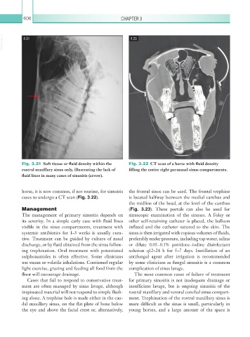
Fig. 3.21 Soft tissue or fluid density within the Fig. 3.22 CT scan of a horse with fluid density
rostral maxillary sinus only, illustrating the lack of filling the entire right paranasal sinus compartments.
fluid lines in many cases of sinusitis (arrow).
horse, it is now common, if not routine, for sinusitis the frontal sinus can be used. The frontal trephine
cases to undergo a CT scan (Fig. 3.22). is located halfway between the medial canthus and
the midline of the head, at the level of the canthus
Management (Fig. 3.23). These portals can also be used for
The management of primary sinusitis depends on sinuscopic examination of the sinuses. A Foley or
its severity. In a simple early case with fluid lines other self-retaining catheter is placed, the balloon
visible in the sinus compartments, treatment with inflated and the catheter sutured to the skin. The
systemic antibiotics for 1–3 weeks is usually cura- sinus is then irrigated with copious volumes of fluids,
tive. Treatment can be guided by culture of nasal preferably under pressure, including tap water, saline
discharge, or by fluid obtained from the sinus follow- or dilute 0.05–0.1% povidone–iodine disinfectant
ing trephination. Oral treatment with potentiated solution q12–24 h for 5–7 days. Instillation of an
sulphonamides is often effective. Some clinicians antifungal agent after irrigation is recommended
use steam or volatile inhalations. Continued regular by some clinicians as fungal sinusitis is a common
light exercise, grazing and feeding all food from the complication of sinus lavage.
floor will encourage drainage. The most common cause of failure of treatment
Cases that fail to respond to conservative treat- for primary sinusitis is not inadequate drainage or
ment are often managed by sinus lavage, although insufficient lavage, but is ongoing sinusitis of the
inspissated material will not respond to simple flush- rostral maxillary and ventral conchal sinus compart-
ing alone. A trephine hole is made either in the cau- ment. Trephination of the rostral maxillary sinus is
dal maxillary sinus, on the flat plate of bone below more difficult as the sinus is small, particularly in
the eye and above the facial crest or, alternatively, young horses, and a large amount of the space is