
Page 218 - Anatomy and Physiology of Farm Animals, 8th Edition
P. 218
Anatomy of the Nervous System / 203
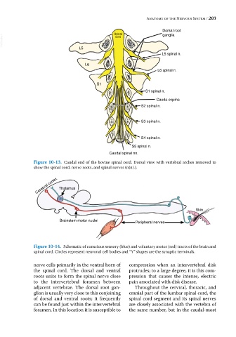
Dorsal root
Spinal
ganglia
VetBooks.ir cord
L5
L5 spinal n.
L6
L6 spinal n.
S1
S1 spinal n.
Cauda equina
S2 spinal n.
S3 spinal n.
S4 spinal n.
S5 spinal n.
Caudal spinal nn.
Figure 10-13. Caudal end of the bovine spinal cord. Dorsal view with vertebral arches removed to
show the spinal cord, nerve roots, and spinal nerves (n(n).).
Cerebral cortex Thalamus
Skin
Brainstem motor nuclei Peripheral nerves
Figure 10-14. Schematic of conscious sensory (blue) and voluntary motor (red) tracts of the brain and
spinal cord. Circles represent neuronal cell bodies and “Y” shapes are the synaptic terminals.
nerve cells primarily in the ventral horn of compression when an intervertebral disk
the spinal cord. The dorsal and ventral protrudes; to a large degree, it is this com-
roots unite to form the spinal nerve close pression that causes the intense, electric
to the intervertebral foramen between pain associated with disk disease.
adjacent vertebrae. The dorsal root gan- Throughout the cervical, thoracic, and
glion is usually very close to this conjoining cranial part of the lumbar spinal cord, the
of dorsal and ventral roots; it frequently spinal cord segment and its spinal nerves
can be found just within the intervertebral are closely associated with the vertebra of
foramen. In this location it is susceptible to the same number, but in the caudal‐most