
Page 216 - Anatomy and Physiology of Farm Animals, 8th Edition
P. 216
Anatomy of the Nervous System / 201
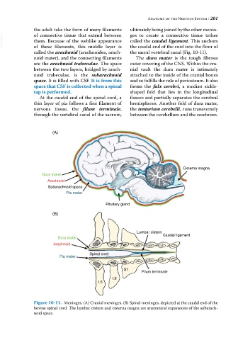
the adult take the form of many filaments ultimately being joined by the other menin-
ges to create a connective tissue tether
of connective tissue that extend between
VetBooks.ir them. Because of the weblike appearance called the caudal ligament. This anchors
the caudal end of the cord into the floor of
of these filaments, this middle layer is
called the arachnoid (arachnoidea, arach- the sacral vertebral canal (Fig. 10‐11).
noid mater), and the connecting filaments The dura mater is the tough fibrous
are the arachnoid trabeculae. The space outer covering of the CNS. Within the cra-
between the two layers, bridged by arach- nial vault the dura mater is intimately
noid trabeculae, is the subarachnoid attached to the inside of the cranial bones
space. It is filled with CSF. It is from this and so fulfills the role of periosteum. It also
space that CSF is collected when a spinal forms the falx cerebri, a median sickle‐
tap is performed. shaped fold that lies in the longitudinal
At the caudal end of the spinal cord, a fissure and partially separates the cerebral
thin layer of pia follows a fine filament of hemispheres. Another fold of dura mater,
nervous tissue, the filum terminale, the tentorium cerebelli, runs transversely
through the vertebral canal of the sacrum, between the cerebellum and the cerebrum.
(A)
Cisterna magna
Dura mater
Arachnoid
Subarachnoid space
Pia mater
Pituitary gland
(B)
Lumbar cistern
Caudal ligament
Dura mater
Arachnoid
Spinal cord
Pia mater
S1
Filum terminale
L6
L5
Figure 10-11. Meninges. (A) Cranial meninges. (B) Spinal meninges, depicted at the caudal end of the
bovine spinal cord. The lumbar cistern and cisterna magna are anatomical expansions of the subarach-
noid space.