
Page 290 - Anatomy and Physiology of Farm Animals, 8th Edition
P. 290
The Integument / 275
Cells in the stratum basale undergo weight‐bearing structures, such as foot-
pads and hoofs. The interface between
mitotic division, which pushes the more
VetBooks.ir superficial layers still farther from the epidermal pegs and dermal papillae pro-
blood vessels in the underlying dermis. As
vides increased surface area for formation
distance from nutrients increases, the cells of a strong junction between these two
flatten and die, leaving a dense mat of their layers. A blister is a local disruption of this
primary constituent, the fibrous protein association between layers, usually due to
keratin. The drying and hardening of the repeated trauma or thermal injury.
superficial cells, a process called both Arteries, veins, capillaries, and lymphat-
keratinization and cornification, renders ics of the skin are contained in the dermis.
the surface of the skin tough and resistant Sensory nerve fibers, in addition to supply-
to drying. As the stratum basale continu- ing the dermis, may extend a short distance
ously adds cells to overlying layers, the into the epidermis. Sympathetic nerves pro-
stratum corneum flakes off and is replaced. vide motor innervation to blood vessels,
The rate at which this occurs can be influ- glands, and arrector pili muscles of hair fol-
enced by trauma or disease processes. A licles in the dermis. These structures do not
callus is a local increase in thickness in receive parasympathetic innervation.
response to continuous trauma. Color of skin is due to the pigment gran-
ules generated in the cytoplasm of the resi-
dent pigment cells, melanocytes. These
Dermis cells in the stratum basale produce the
pigment, melanin, which is brown, yellow-
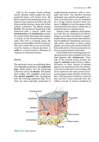
The epidermis forms an undulating sheet ish‐brown, or black. Packets of melanin
with fingerlike projections, the epidermal pigment are manufactured by the melano-
pegs, which project into the underlying cytes and transferred to surrounding cells
connective tissue, the dermis. The dermis of the epidermis; the same process incor-
bears ridges and nipplelike projections porates pigment into cells that cornify into
(the dermal papillae) that interdigitate hairs. The expression of different colors in
with the overlying epidermis (Fig. 14‐2); skin and hair comes primarily from the
these are most especially prominent in relative amount of melanin produced in
Sebaceous gland
Sweat gland
EPIDERMIS
DERMIS
Hair follicle
Nerve endings
HYPODERMIS
Blood vessels
Fat cells
Figure 14-2. Skin anatomy.