
Page 373 - Anatomy and Physiology of Farm Animals, 8th Edition
P. 373
358 / Anatomy and Physiology of Farm Animals
(A) (B) (C)
VetBooks.ir a a c a c
d
d b b d
d d d d b b d d b b
d d d
d d
d
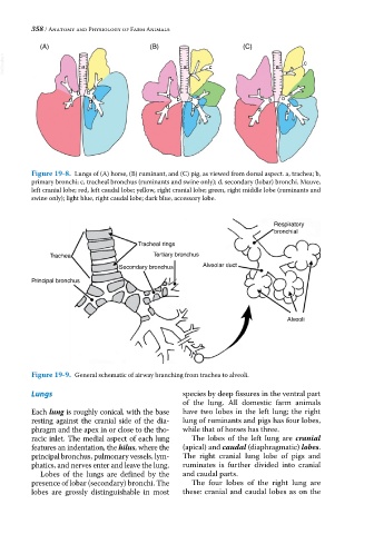
Figure 19-8. Lungs of (A) horse, (B) ruminant, and (C) pig, as viewed from dorsal aspect. a, trachea; b,
primary bronchi; c, tracheal bronchus (ruminants and swine only); d, secondary (lobar) bronchi. Mauve,
left cranial lobe; red, left caudal lobe; yellow, right cranial lobe; green, right middle lobe (ruminants and
swine only); light blue, right caudal lobe; dark blue, accessory lobe.
Respiratory
bronchial
Tracheal rings
Trachea Tertiary bronchus
Secondary bronchus Alveolar duct
Principal bronchus
Alveoli
Figure 19-9. General schematic of airway branching from trachea to alveoli.
Lungs species by deep fissures in the ventral part
of the lung. All domestic farm animals
Each lung is roughly conical, with the base have two lobes in the left lung; the right
resting against the cranial side of the dia- lung of ruminants and pigs has four lobes,
phragm and the apex in or close to the tho- while that of horses has three.
racic inlet. The medial aspect of each lung The lobes of the left lung are cranial
features an indentation, the hilus, where the (apical) and caudal (diaphragmatic) lobes.
principal bronchus, pulmonary vessels, lym- The right cranial lung lobe of pigs and
phatics, and nerves enter and leave the lung. ruminates is further divided into cranial
Lobes of the lungs are defined by the and caudal parts.
presence of lobar (secondary) bronchi. The The four lobes of the right lung are
lobes are grossly distinguishable in most these: cranial and caudal lobes as on the