
Page 375 - Anatomy and Physiology of Farm Animals, 8th Edition
P. 375
360 / Anatomy and Physiology of Farm Animals
Pleura For this reason infections or air in one
pleural space may stay unilaterally
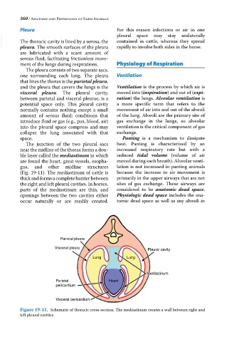
VetBooks.ir The thoracic cavity is lined by a serosa, the contained in cattle, whereas they spread
rapidly to involve both sides in the horse.
pleura. The smooth surfaces of the pleura
are lubricated with a scant amount of
serous fluid, facilitating frictionless move-
ment of the lungs during respirations. Physiology of Respiration
The pleura consists of two separate sacs,
one surrounding each lung. The pleura Ventilation
that lines the thorax is the parietal pleura,
and the pleura that covers the lungs is the Ventilation is the process by which air is
visceral pleura. The pleural cavity, moved into (inspiration) and out of (expi-
between parietal and visceral pleurae, is a ration) the lungs. Alveolar ventilation is
potential space only. This pleural cavity a more specific term that refers to the
normally contains nothing except a small movement of air into and out of the alveoli
amount of serous fluid; conditions that of the lung. Alveoli are the primary site of
introduce fluid or gas (e.g., pus, blood, air) gas exchange in the lungs, so alveolar
into the pleural space compress and may ventilation is the critical component of gas
collapse the lung associated with that exchange.
space. Panting is a mechanism to dissipate
The junction of the two pleural sacs heat. Panting is characterized by an
near the midline of the thorax forms a dou- increased respiratory rate but with a
ble layer called the mediastinum in which reduced tidal volume (volume of air
are found the heart, great vessels, esopha- moved during each breath). Alveolar venti-
gus, and other midline structures lation is not increased in panting animals
(Fig. 19‐11). The mediastinum of cattle is because the increase in air movement is
thick and forms a complete barrier between primarily in the upper airways that are not
the right and left pleural cavities. In horses, sites of gas exchange. These airways are
parts of the mediastinum are thin, and considered to be anatomic dead space.
openings between the two cavities either Physiologic dead space includes the ana-
occur naturally or are readily created. tomic dead space as well as any alveoli in
Parietal pleura
Visceral pleura Pleural cavity
Lung Lung
Mediastinum
Mediastinum
Parietal Heart
pericardium
Visceral pericardium
Figure 19-11. Schematic of thoracic cross‐section. The mediastinum creates a wall between right and
left pleural cavities