
Page 420 - Anatomy and Physiology of Farm Animals, 8th Edition
P. 420
Physiology of Digestion / 405
cells secrete the enzymes, and cells that line protected from an acidic solution by a
thick layer of mucus, as is the stomach.
ducts in the pancreas secrete the sodium
VetBooks.ir bicarbonate (Fig. 21‐7). These ducts empty The higher pH is also better for the action
of the pancreatic digestive enzymes. The
into one or two pancreatic ducts, which
empty into the duodenum (see Fig. 20‐19). major stimulus for bicarbonate secretion is
The sodium bicarbonate raises the pH the hormone secretin from the small intes-
of the chyme entering from the stomach. tinal mucosa. Secretin secretion increases
The small intestinal epithelium is not in response to the acid chyme entering
from the stomach.
Pancreatic proteolytic enzymes include
(A) trypsin and chymotrypsin. Similar to pep-
Food bolus
sin in the stomach, these are secreted as
inactive precursors, trypsinogen and
chymotrypsinogen.
Trypsinogen is activated by an enzyme,
(B) enterokinase, a component of the luminal
cell membranes of small intestinal cells
(enterocytes). Trypsin can activate chymo-
trypsinogen and more trypsinogen. The
ultimate end products of protein digestion
are amino acids, but the pancreatic proteo-
(C) lytic enzymes may stop digestion when the
peptides reach a length of two or more
amino acids. If this occurs, peptidases
associated with enterocyte cell membranes
can complete hydrolysis of the peptides to
individual amino acids for absorption.
Figure 21-6. Segmentation pattern of gastroin-
testinal motility. The same segment of intestine is Unlike the proteolytic enzymes, pan-
viewed at three different sequential times (A, B, creatic amylase and lipase are in the
and C). Regions of contractions at each time are active forms when secreted from the
identified by arrows. pancreas. Amylase digests starches to
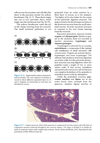
Figure 21-7. Equine pancreas. Most of the pancreas is composed of exocrine acinar cells with islets of
Langerhans (arrows) scattered throughout the exocrine regions. The organ is divided in to lobules by
septa of connective tissue with complex duct systems. Source: Bacha and Bacha, 2012. Reproduced with
permission of John Wiley & Sons, Inc.