
Page 83 - Clinical Small Animal Internal Medicine
P. 83
7 Pituitary-Dependent Hyperadrenocorticism in Dogs and Cats 51
Corticotroph and Melanotroph Cell Marker: Tpit
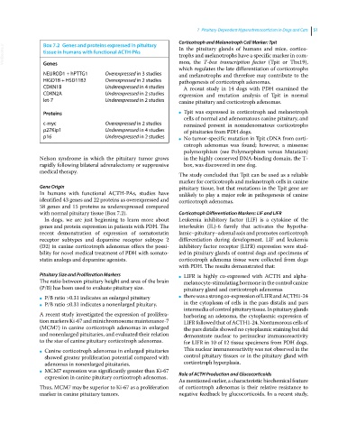
Box 7.2 Genes and proteins expressed in pituitary
VetBooks.ir tissue in humans with functional ACTH‐PAs trophs and melanotrophs have a specific marker in com
In the pituitary glands of humans and mice, cortico
mon, the T‐box transcription factor (Tpit or Tbx19),
Genes
which regulates the late differentiation of corticotrophs
NEUROD1 + hPTTG1 Overexpressed in 3 studies and melanotrophs and therefore may contribute to the
HIGD1B + HSD11B2 Overexpressed in 2 studies pathogenesis of corticotroph adenomas.
CDKN1B Underexpressed in 4 studies A recent study in 14 dogs with PDH examined the
CDKN2A Underexpressed in 2 studies expression and mutation analysis of Tpit in normal
let‐7 Underexpressed in 2 studies canine pituitary and corticotroph adenomas.
Proteins ● Tpit was expressed in corticotroph and melanotroph
cells of normal and adenomatous canine pituitary, and
c‐myc Overexpressed in 2 studies remained present in nonadenomatous corticotrophs
p27Kip1 Underexpressed in 4 studies of pituitaries from PDH dogs.
p16 Underexpressed in 2 studies No tumor‐specific mutation in Tpit cDNA from corti
●
cotroph adenomas was found; however, a missense
polymorphism (see Polymorphism versus Mutation)
Nelson syndrome in which the pituitary tumor grows in the highly conserved DNA‐binding domain, the T‐
rapidly following bilateral adrenalectomy or suppressive box, was discovered in one dog.
medical therapy.
The study concluded that Tpit can be used as a reliable
marker for corticotroph and melanotroph cells in canine
Gene Origin pituitary tissue, but that mutations in the Tpit gene are
In humans with functional ACTH‐PAs, studies have unlikely to play a major role in pathogenesis of canine
identified 43 genes and 22 proteins as overexpressed and corticotroph adenomas.
58 genes and 15 proteins as underexpressed compared
with normal pituitary tissue (Box 7.2). Corticotroph Differentiation Markers: LIF and LIFR
In dogs, we are just beginning to learn more about Leukemia inhibitory factor (LIF) is a cytokine of the
genes and protein expression in patients with PDH. The interleukin (IL)‐6 family that activates the hypotha
recent demonstration of expression of somatostatin lamic–pituitary–adrenal axis and promotes corticotroph
receptor subtypes and dopamine receptor subtype 2 differentiation during development. LIF and leukemia
(D2) in canine corticotroph adenomas offers the possi inhibitory factor receptor (LIFR) expression were stud
bility for novel medical treatment of PDH with somato ied in pituitary glands of control dogs and specimens of
statin analogs and dopamine agonists. corticotroph adenoma tissue were collected from dogs
with PDH. The results demonstrated that:
Pituitary Size and Proliferation Markers ● LIFR is highly co‐expressed with ACTH and alpha‐
The ratio between pituitary height and area of the brain melanocyte‐stimulating hormone in the control canine
(P/B) has been used to evaluate pituitary size. pituitary gland and corticotroph adenomas
P/B ratio >0.31 indicates an enlarged pituitary ● there was a strong co‐expression of LIFR and ACTH1‐24
●
P/B ratio ≤0.31 indicates a nonenlarged pituitary. in the cytoplasm of cells in the pars distalis and pars
●
intermedia of control pituitary tissue. In pituitary glands
A recent study investigated the expression of prolifera harboring an adenoma, the cytoplasmic expression of
tion markers Ki‐67 and minichromosome maintenance‐7 LIFR followed that of ACTH1‐24. Nontumorous cells of
(MCM7) in canine corticotroph adenomas in enlarged the pars distalis showed no cytoplasmic staining but did
and nonenlarged pituitaries, and evaluated their relation demonstrate nuclear to perinuclear immunoreactivity
to the size of canine pituitary corticotroph adenomas. for LIFR in 10 of 12 tissue specimens from PDH dogs.
Canine corticotroph adenomas in enlarged pituitaries This nuclear immunoreactivity was not observed in the
●
showed greater proliferation potential compared with control pituitary tissues or in the pituitary gland with
adenomas in nonenlarged pituitaries. corticotroph hyperplasia.
MCM7 expression was significantly greater than Ki‐67
● Role of ACTH Production and Glucocorticoids
expression in canine pituitary corticotroph adenomas.
As mentioned earlier, a characteristic biochemical feature
Thus, MCM7 may be superior to Ki‐67 as a proliferation of corticotroph adenomas is their relative resistance to
marker in canine pituitary tumors. negative feedback by glucocorticoids. In a recent study,