
Page 161 - BSAVA Manual of Canine and Feline Head, Neck and Thoracic Surgery, 2nd Edition
P. 161
BSAVA Manual of Canine and Feline Head, Neck and Thoracic Surgery
VetBooks.ir
(a) (b)
(c) (d)
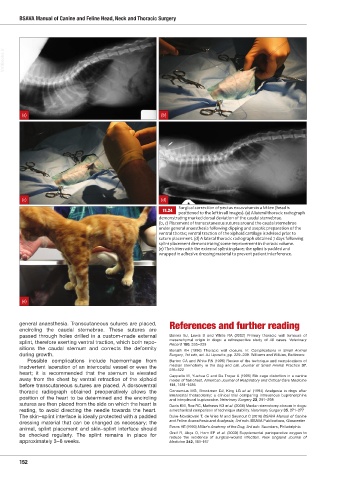
Surgical correction of pectus excavatum in a kitten (head is
11.24 positioned to the left in all images). (a) A lateral thoracic radiograph
demonstrating marked dorsal deviation of the caudal sternebrae.
(b, c) Placement of transcutaneous sutures around the caudal sternebrae
under general anaesthesia following clipping and aseptic preparation of the
ventral thora ventral traction of the iphoid cartilage is advised prior to
suture placement. (d) A lateral thoracic radiograph obtained 7 days following
splint placement demonstrating some improvement in thoracic volume.
e The kitten ith the e ternal splint in place the splint is padded and
wrapped in adhesive dressing material to prevent patient interference.
(e)
general anaesthesia. Transcutaneous sutures are placed, References and further reading
encircling the caudal sternebrae. These sutures are
passed through holes drilled in a custom-made external Baines SJ, Lewis S and White RA (2002) Primary thoracic wall tumours of
mesenchymal origin in dogs: a retrospective study of 46 cases. Veterinary
splint, therefore exerting ventral traction, which both repo-
Record 150, 335–339
sitions the caudal sternum and corrects the deformity
Bonath KH (1996) Thoracic wall closure. In: Complications in Small Animal
during growth. Surgery, 1st edn, ed. AJ Lipowitz, pp. 229–239. Williams and Wilkins, Baltimore
Possible complications include haemorrhage from Burton CA and White RN (1996) Review of the technique and complications of
inadvertent laceration of an intercostal vessel or even the median sternotomy in the dog and cat. Journal of Small Animal Practice 37,
516–522
heart; it is recommended that the sternum is elevated
Cappello M, Yuehua C and De Troyer A (1995) Rib cage distortion in a canine
away from the chest by ventral retraction of the xiphoid model of flail chest. American Journal of Respiratory and Critical Care Medicine
before transcutaneous sutures are placed. A dorsoventral 151, 1481–1485
thoracic radiograph obtained preoperatively allows the Conzemius MG, Brockman DJ, King LG et al. (1994) Analgesia in dogs after
intercostal thoracotomy: a clinical trial comparing intravenous buprenorphine
position of the heart to be determined and the encircling
and interpleural bupivacaine. Veterinary Surgery 23, 291–298
sutures are then placed from the side on which the heart is Davis KM, Roe RC, Mathews KG et al. (2006) Median sternotomy closure in dogs:
resting, to avoid directing the needle towards the heart. a mechanical comparison of technique stability. Veterinary Surgery 35, 271–277
The skin–splint interface is ideally protected with a padded Duke-Novakovski T, de Vries M and Seymour C (2016) BSAVA Manual of Canine
dressing material that can be changed as necessary; the and Feline Anaesthesia and Analgesia, 3rd edn. BSAVA Publications, Gloucester
animal, splint placement and skin–splint interface should Evans HE (1993) Miller’s Anatomy of the Dog, 3rd edn. Saunders, Philadelphia
Greif R, Akça O, Horn EP et al. (2000) Supplemental perioperative oxygen to
be checked regularly. The splint remains in place for
reduce the incidence of surgical-wound infection. New England Journal of
approximately 3–6 weeks. Medicine 342, 161–167
152
Ch11 HNT.indd 152 31/08/2018 11:52