
Page 145 - Avian Virology: Current Research and Future Trends
P. 145
138 | Liu et al.
Figure 5.5 Genome organization of infectious bronchitis virus (IBV). The IBV viral RNA genome is approximately 27.6 kilobases (kb) long and
is arranged in an invariant order of 5′-rep 1a-rep 1b-S-3a-3b-E-M-5a-5b-N-3’.
Figure 5. Genome organization of infectious bronchitis virus (IBV). The IBV viral RNA genome is approximately 27.6 kilobases (kb) long and is arranged
in an invariant order of 5’-rep 1a-rep 1b-S-3a-3b-E-M-5a-5b-N-3’.
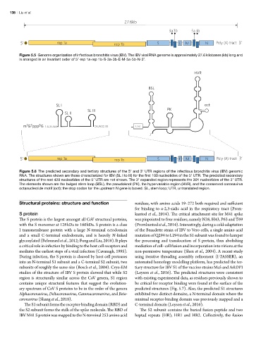
Figure 6. The predicted secondary and tertiary structures of the 5’ and 3’ UTR regions of the infectious bronchitis virus (IBV) genomic RNA. The
Figure 5.6 The predicted secondary and tertiary structures of the 5′ and 3′ UTR regions of the infectious bronchitis virus (IBV) genomic
structures shown are those characterized for IBV (SL I to III) for the first 100 nucleotides of the 5’ UTR. The predicted secondary structures of the rest 428
RNA. The structures shown are those characterized for IBV (SL I to III) for the first 100 nucleotides of the 5′ UTR. The predicted secondary
nucleotides of the 5’ UTR are not shown. The 3’ expanded region represents the 301 nucleotides of the 3’ UTR. The elements shown are the bulged stem loop
structures of the rest 428 nucleotides of the 5′ UTR are not shown. The 3′ expanded region represents the 301 nucleotides of the 3′ UTR.
(BSL), the pseudoknot (PK), the hypervariable region (HVR), and the conserved coronavirus octanucleotide motif (oct); the stop codon for the upstream N gene
The elements shown are the bulged stem loop (BSL), the pseudoknot (PK), the hypervariable region (HVR), and the conserved coronavirus
is boxed. UTR, untranslated region; SL, stem loop.
octanucleotide motif (oct); the stop codon for the upstream N gene is boxed. SL, stem loop; UTR, untranslated region.
Structural proteins: structure and function residues, with amino acids 19–272 both required and sufficient
for binding to α-2,3-sialic acid in the respiratory tract (Prom-
S protein kuntod et al., 2014). The critical attachment site for M41 spike
The S protein is the largest amongst all CoV structural proteins, was pinpointed to four residues, namely N38, H43, P63 and T69
with the S monomer at 128 kDa to 160 kDa. S protein is a class (Promkuntod et al., 2014). Interestingly, during a cold-adaptation
I transmembrane protein with a large N-terminal ectodomain of the Beaudette strain of IBV to Vero cells, a single amino acid
and a small C-terminal endodomain, and is heavily N-linked mutation of Q294 to L294 in the S1 subunit was found to hamper
glycosylated (Belouzard et al., 2012; Fung and Liu, 2018). It plays the processing and translocation of S protein, thus abolishing
a critical role in infection by binding to the host cell receptors and mediation of cell–cell fusion and incorporation into virions at the
mediates the earliest steps of a viral infection (Cavanagh, 1995). non-permissive temperature (Shen et al., 2004). A recent study
During infection, the S protein is cleaved by host cell proteases using iterative threading assembly refinement (I-TASSER), an
into an N-terminal S1 subunit and a C-terminal S2 subunit, two automated homology modelling platform, has predicted the ter-
subunits of roughly the same size (Bosch et al., 2004). Cryo-EM tiary structure for IBV S1 of the vaccine strains Ma5 and ArkDPI
studies of the structure of IBV S protein showed that while S2 (Leyson et al., 2016). The predicted structures were consistent
region is structurally similar across the CoV genera, S1 region with existing experimental data, as residues previously shown to
contains unique structural features that suggest the evolution- be critical for receptor binding were found at the surface of the
ary spectrum of CoV S proteins to be in the order of the genera predicted structures (Fig. 5.7). Also, the predicted S1 structures
Alphacoronavirus, Deltacoronavirus, Gammacoronavirus, and Beta- exhibited two distinct domains, a N-terminal domain where the
coronavirus (Shang et al., 2018). minimal receptor-binding domain was previously mapped and a
The S1 subunit forms the receptor-binding domain (RBD) and C-terminal domain (Leyson et al., 2016).
the S2 subunit forms the stalk of the spike molecule. The RBD of The S2 subunit contains the buried fusion peptide and two
IBV M41 S protein was mapped to the N-terminal 253 amino acid heptad repeats (HR), HR1 and HR2. Collectively, the fusion