
Page 146 - Avian Virology: Current Research and Future Trends
P. 146
Infectious Bronchitis Virus | 139
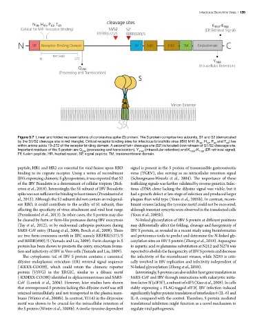
Figure 5.7 Linear and folded representations of coronavirus spike (S) protein. The S protein comprise two subunits, S1 and S2 (demarcated
by the S1/S2 cleavage site in red triangle). Critical receptor binding sites for infectious bronchitis virus (IBV) M41 (N , H , P and T ) lies
38 43 63 69
within amino acids 19–272 of the receptor binding domain. A second furin cleavage site (S2’) is located downstream of S1/S2 cleavage site.
Important residues of the S protein are Q (processing and translocation), Y (intracellular retention) and K -K (ER retrieval signal).
294 1143 1159 1160
FP, fusion peptide; HR, heptad repeat; SP, signal peptide; TM, transmembrane domain.
peptide, HR1 and HR2 are essential for viral fusion upon RBD signal is present in the S protein of transmissible gastroenteritis
binding to its cognate receptor. Using a series of recombinant virus (TGEV), also serving as an intracellular retention signal
IBVs expressing chimeric S glycoproteins, it was reported that S2 (Schwegmann-Wessels et al., 2004). The importance of these
of the IBV Beaudette is a determinant of cellular tropism (Bick- trafficking signals was further validated by reverse genetics. Infec-
erton et al., 2018). Interestingly, the S1 subunit of IBV Beaudette tious cDNA clone lacking the dilysine signal was viable, but it
spike was not sufficient for binding to host tissues (Promkuntod et had a growth defect at late stage of infection and produced larger
al., 2013). Although the S2 subunit did not contain an independ- plaques than wild type (Youn et al., 2005b). In contrast, recom-
ent RBD, it could contribute to the avidity of S1 subunit, thus binant viruses lacking the tyrosine motif could not be recovered,
affecting the specificity of virus attachment and viral host range although transient syncytia were observed in the transfected cells
(Promkuntod et al., 2013). In other cases, the S protein may also (Youn et al., 2005b).
be cleaved by furin or furin-like proteases during IBV exocytosis N-linked glycosylation of IBV S protein at different positions
(Tay et al., 2012), or by endosomal cathepsin proteases during may differentially affect the folding, cleavage and fusogenicity of
SARS-CoV entry (Huang et al., 2006; Bosch et al., 2008). There IBV S protein, as revealed in a recent study using bioinformatics
are two furin consensus motifs in IBV, namely RRFRR(537)/S and proteomics tools to predict and determine the N-linked gly-
and RRRR(690)/S (Yamada and Liu, 2009). Furin cleavage in S cosylation sites on IBV S protein (Zheng et al., 2018). Asparagine
protein has been shown to promote the entry, syncytium forma- to aspartic acid or glutamine substitution at N212 and N276 was
tion and infectivity of IBV in Vero cells (Yamada and Liu, 2009). reported to abolish the fusogenicity of IBV S protein and decrease
The cytoplasmic tail of IBV S protein contains a canonical the infectivity of the recombinant viruses, while N283 is criti-
dilysine endoplasmic reticulum (ER) retrieval signal sequence cally involved in IBV replication and infectivity independent of
(-KKXX-COOH), which could retain the chimeric reporter N-linked glycosylation (Zheng et al., 2018).
protein (VSVG) in the ERGIC, similar to a dibasic motif Interestingly, S protein can also inhibit host gene translation in
(-KXHXX-COOH) identified in alphacoronaviruses and SARS- SARS-CoV and IBV through interactions with eukaryotic initia-
CoV (Lontok et al., 2004). However, later studies have shown tion factor 3f (eIF3F), a subunit of eIF3 (Xiao et al., 2008). In cells
that overexpressed S proteins lacking this dilysine motif was still stably expressing a FLAG-tagged eIF3f, IBV infection induced
retained intracellularly and not transported to the plasma mem- significantly higher protein translation of interleukin 6 (IL-6) and
brane (Winter et al., 2008b). In contrast, Y1143 in the dityrosine IL-8, compared with the control. Therefore, S protein mediated
motif was shown to be crucial for the intracellular retention of translational inhibition might function as a novel mechanism to
the S protein (Winter et al., 2008b). A similar tyrosine dependent regulate viral pathogenesis.