
Page 205 - Avian Virology: Current Research and Future Trends
P. 205
198 | Kibenge et al.
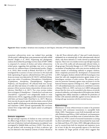
Figure 6.5 Winter mortality in American crow (courtesy of Jordi Segers, University of Prince Edward Island, Canada).
reassortant orthoreovirus strain was isolated from partridge 1 day old. Those infected orally at 7 days and 3 weeks showed a
(Perdix perdix) suffering from acute pneumonia and infra-orbital substantial rise in intestinal IgA. In the subcutaneously infected
sinusitis (Kugler et al., 2016). Sequencing and phylogenetic chicks, only those infected at 3 weeks showed an intestinal IgA
analyses showed that the partridge reovirus strain (D1007/2008) response. There were very similar reovirus-specific IgG responses
is composed of a mixture of chicken and turkey orthoreovirus in the sera of the orally and subcutaneously infected chicks, in
related genes, suggesting that partridges may serve as natural all age-groups. Previously, Meanger et al. (1997) had shown that
reservoirs for orthoreoviruses of domesticated poultry (Kugler vaccination of breeding hens with avian reovirus resulted in the
et al., 2016). Most recently in Poland, Styś-Fijoł et al. (2017) passive transfer of neutralizing antibody to progeny chickens,
investigated the occurrence of avian reoviruses in 192 dead wild which completely prevented the development of tenosynovitis
birds representing 32 species collected between 2014 and 2016. in 80% of progeny chickens infected with the homologous virus
Avian reoviruses were detected in 58 (30.2%) wild birds belong- strain but with only marginal protection against strains of two
ing to nine orders (Ciconiiformes, Pelecaniformes, Columbiformes, heterologous serotypes of avian reovirus. The primary mecha-
Accipitriformes, Anseriformes, Charadariiformes, Strigiformes, nism for protective immunity against avian reoviruses is via the
Piciformes, and Passeriformes). All collected birds were necrop- humoral immune response (Kibenge et al., 1987). A suppression
sied, and often had lesions of swollen liver and spleen with the of T-cell mediated immunity was shown to contribute to severity
presence of liver necrosis; lesions characteristic of avian reovirus of disease (Hill et al., 1989). van Loon et al. (2003) subsequently
infection (Styś-Fijoł et al., 2017). Two avian reovirus isolates showed that the virus can be controlled in the absence of actively
from rock pigeon (Columbiformes – Columba livia) and mute swan produced antibodies, and independent of B lymphocytes, further
(Anseriformes – Cygnusolor) were antigenically similar to avian suggesting that cellular immunity is sufficient for protection of
orthoreovirus S1133, suggesting possible transmission between broilers with maternal antibodies against reovirus infection fol-
wild birds and farmed birds (Styś-Fijoł et al., 2017). However, lowing early age vaccination with live reoviral vaccine.
there may also be species-specific reovirus strains, some of which
may be capable of inducing cross-infections among species. Jones
and Guneratne (1984) showed experimentally that reoviruses Epizootiology
isolated from wedge-tailed eagle (Aquila audax) are pathogenic Maintenance of avian orthoreoviruses in nature is by persistent
for chickens. infections with continuous transmission to susceptible birds
(Stott, 1999). Transmission occurs both horizontally and verti-
cally (Robertson and Wilcox, 1986) with faecal–oral transmission
Immune responses being considered the most likely route of natural infection (Mac-
The IgA and IgG responses of chickens to avian reoviruses and donald et al., 1978). Lateral or horizontal transmission occurring
the effects of age of chicks (1-day-old, 7-day-old and 3-week-old) via direct and indirect contact was demonstrated by many stud-
and route of infection (oral and subcutaneous inoculation) were ies (Sahu and Olson, 1975; Stott, 1999). Although orthoreovirus
investigated by Mukiibi-Muka and Jones (1999). Virus titres in may be excreted from both the intestinal and respiratory tracts
the gut declined with increasing age of chick at infection. IgA for at least 10 dpi, the virus generally appears to be shed from
was not detected in the intestinal contents of chicks infected at the intestine for longer periods of time which suggests that