
Page 200 - Avian Virology: Current Research and Future Trends
P. 200
Avian Reovirus | 193
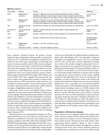
Table 6.2 Continued
Virus isolate Disease Source Reference
ERS-1 Malabsorption Isolated in 1998 from young commercially reared broilers flocks in Poland van Loon et al.
syndrome manifesting difficulty in walking, high mortality, hepatic necrosis and pericarditis. (2001)
The virus was isolated from liver, kidney, thymus, caecal tonsils, spleen, and heart
ERS-2 Malabsorption Isolated in 1999 from young commercially reared broilers flocks in Poland van Loon et al.
syndrome manifesting difficulty in walking, high mortality, hepatic necrosis, and pericarditis. (2001)
The virus was isolated from liver, kidney, thymus, caecal tonsils, spleen and heart
VAA Tenosynovitis/viral Isolated from a chickens with ruptured gastrocnemius tendons, and viral arthritis Jones et al. (1975)
arthritis
724, 846, 847, Tenosynovitis/viral Isolated in western Australia from broiler breeder chickens affected with Kibenge et al.
848 arthritis tenosynovitis (1982)
1091 Malabsorption Isolated in Australia from broiler chickens affected by runting/stunting syndrome Pass et al. (1982)
syndrome
RAM-1 None Isolated in Australia from a chicken kidney cell culture Mustaffa-Babjee
and Spradbrow
(1971)
OS161 Malabsorption Isolated in 1970 from a chicken in Japan Shen et al. (2007)
syndrome
T6 Respiratory disease Isolated in 1970 from chickens in Taiwan Shen et al. (2007)
firmer, adhesions developed between the tendons, synovial of the synovial lining cells and a diffuse infiltrate of lymphocytes,
sheath and skin, rendering the tendons partially non-functional plasma cells, macrophages, and a few heterophils. Clumps of
(Johnson, 1972). A yellowish-brown gelatinous exudate between heterophils and desquamated synovial cells were occasionally
tendons in the swollen legs, varying degrees of thickening and present in the synovial spaces. Chronic inflammatory changes
fusion of tendons, and pitted erosions of the articular cartilage of were evident in the articular cartilage by replacement of the car-
the hock joints were observed by 12 weeks PI (Jones and Kibenge, tilage by connective tissue (pannus) that was grossly evident as
1984; Jones and Georgiou, 1985). Acute avian arthritis was char- pitting of the articular surface (Gouvea and Schnitzer, 1982a). At
acterized by an initial inflammatory response in the joints that 7.5 weeks PI, van der Heide et al. (1974) observed chronic fibro-
progressed in many cases to pannus formation, erosion of under- sis of the tendon sheaths with fibrous connective tissue invading
lying cartilage, and ultimately fibrosis (Stott, 1999). Johnson and and replacing the normal architecture of the tendon, resulting
van der Heide (1971) have suggested that tenosynovitis may in ankylosis and immobility. At 33 weeks PI, mononuclear cell
lead to rupture of the gastrocnemius tendons in mature broiler infiltration was still an inflammatory lesion in the sheaths and
breeders. Johnson (1972) observed the tendons started to tear tendons. Heterophils were still prominent in some areas and large
creating haemorrhage at tendon muscle junction that proceeded lymphoid foci were occasionally seen at the tendon periphery
to rupture with time, and Jones and Georgiou (1984) observed (Jones and Onunkwo, 1978). Fibroplasia had also occurred in the
the rupture of digital flexor tendons by 6 weeks PI. tendon sheaths. According to Islam et al. (1990), infection with
Microscopic lesions of acute viral arthritis include oedema, arthrotropic avian orthoreovirus provided evidence of an auto-
coagulation necrosis, and perivascular infiltration of lymphocytes immune reaction. Although lesions were localized to synovial
and macrophages. Thickening of the tendon sheath was caused by structures, the virus was widely distributed in various surround-
reticular cell proliferation, synovial cell hyperplasia, and hyper- ing tissues during the early stage of infection (Menendez et al.,
trophy, infiltrates of heterophils and macrophages, and periostitis. 1975b; Ellis et al., 1983; Kibenge et al., 1985). The ability of avian
Synovial cavities were filled with sloughed synovial and inflamma- orthoreovirus to establish persistent infections may be due to the
tory cells (Stott, 1999). Loose connective tissue surrounding the joint and tendon acting as sequestered sites protecting the virus
sheath was replaced by granulomatous inflammation and fibrous from elimination by the immune system (Jones and Georgiou,
connective tissue. Granulomatous inflammation infiltrated into 1985).
the tendons causing them to adhere firmly to their surrounding
sheath (Johnson, 1972). Respiratory orthoreovirus disease
The chronic disease was characterized by formation of villi on Avian orthoreovirus was originally isolated from chickens with
synovial membranes, an increase in fibrous connective tissue, and acute or chronic respiratory disease (Fahey and Crawley, 1954).
infiltration or proliferation of reticular cells, lymphocytes, mac- Fahey reported the isolation of viruses with identical character-
rophages, and plasma cells (Stott, 1999). Olson and Weiss (1972) istics to those of avian orthoreovirus from ducks (Fahey, 1955)
described the histopathology of birds infected with Fahey–Craw- experiencing chronic respiratory disease as well as turkeys
ley agent via the foot pad. Extensive fibrosis and the presence of (Fahey, 1956) with infectious sinusitis and suggested a common
numerous lymphoid follicles in the digital flexor tendon sheaths aetiological factor as the cause of these respiratory diseases in
was observed 43 days PI. There were hypertrophy and hyperplasia various bird species. Subramanyam and Pomeroy (1960) showed