
Page 147 - Canine Lameness
P. 147
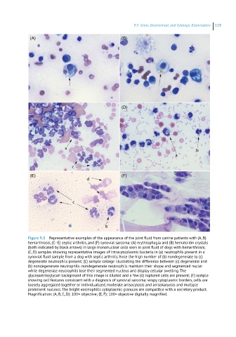
9.3 Groos, BrocheBocas, cand CyraroBo EceBacyBra 119
(A) (B)
(C) (D)
(E) (F)
Figure 9.5 Representative examples of the appearance of the joint fluid from canine patients with (A, B)
hemarthrosis, (C–E) septic arthritis, and (F) synovial sarcoma: (A) erythrophagia and (B) hematoidin crystals
(both indicated by black arrows) in large mononuclear cells seen in joint fluid of dogs with hemarthrosis;
(C, D) samples showing representative images of intracytoplasmic bacteria in (a) neutrophils present in a
synovial fluid sample from a dog with septic arthritis. Note the high number of (b) nondegenerate to (c)
degenerate neutrophils present; (E) sample collage illustrating the difference between (c) degenerate and
(b) nondegenerate neutrophils: nondegenerate neutrophils maintain their shape and segmented nuclei
while degenerate neutrophils lose their segmented nucleus and display cellular swelling. The
glycosaminoglycan background of this image is diluted and a few (d) ruptured cells are present; (F) sample
showing cell features consistent with a diagnosis of synovial sarcoma: wispy cytoplasmic borders, cells are
loosely aggregated together or individualized, moderate anisocytosis and anisokaryosis and multiple
prominent nucleoli. The bright eosinophilic cytoplasmic granules are compatible with a secretory product.
Magnification: (A, B, C, D): 100× objective; (E, F): 100× objective digitally magnified.