
Page 232 - Canine Lameness
P. 232
(A) (B) (C) (D) (E)
(F) (G) (H) (I)
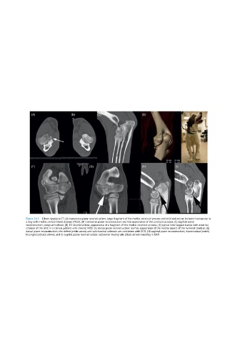
Figure 14.5 Elbow dysplasia CT: (A) transverse plane reconstruction; large fragment of the medial coronoid process and mild radioulnar incisure incongruity in
a dog with medial compartment disease (MCD); (B) transverse plane reconstruction; normal appearance of the coronoid process; (C) sagittal plane
reconstruction; congruent elbow; (D) 3D reconstruction; appearance of a fragment of the medial coronoid process; (E) typical bow-legged stance with external
rotation of the limb in a clinical patient with chronic MCD; (F) dorsal plane reconstruction; normal appearance of the medial aspect of the humeral condyle; (G)
dorsal plane reconstruction; the defect (white arrow) and subchondral sclerosis are consistent with OCD; (H) sagittal plane reconstruction; humeroulnar (notch)
incongruity (black arrow); and (I) sagittal plane reconstruction; radioulnar incongruity (black arrow) resulting in UAP.