
Page 259 - Canine Lameness
P. 259
15.4 Medial Shoulder nstaaility 231
15.4 Medial Shoulder Instability
MSI describes a condition of reduced stability of the shoulder, caused by pathology of the medial
structures of this joint. MSI is the most common type of canine shoulder instability and a frequent
cause of shoulder-related lameness (Cogar et al. 2008). Lateral and multidirectional shoulder insta-
bility has been reported to occur in about 25% of shoulder instability cases (Franklin et al. 2013).
MSI is believed to result from repetitive trauma. Depending on the amount and magnitude of
involved forces, the structures supporting the shoulder can become strained, frayed, disrupted, or
broken down completely. This results in various degrees of instability, including subluxation or
complete luxation (covered in 15.5) of the scapulohumeral joint. As MSI does not always result in
detectable instability, the term “medial shoulder syndrome or disease” has been suggested in the
non-peer-reviewed literature. While the official nomenclature may change in the future, for the
scope of this chapter, MSI will be used throughout.
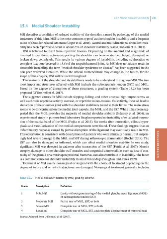
The anatomy of the shoulder and its stabilizers needs to be understood to diagnose MSI. The two
most important structures affected with MSI include the subscapularis tendon (SST) and MGL.
Based on the degree of disruption of these structures, a grading system (Table 15.2) has been
proposed (O’Donnell et al. 2017).
The suggested causes for MSI include slipping, falling, and other unusual high-impact stress, as
well as chronic repetitive activity, overuse, or repetitive micro-trauma. Collectively, these all lead to
abduction of the shoulder joint with the shoulder stabilizers tested to their limits. The main stress
seems to be concentrated on the medial joint capsule, the MGL, and the SST. While it has been sug-
gested that the MGL provides the majority of medial shoulder stability (Sidaway et al. 2004), an
experimental study in purpose bred laboratory Beagles reported no instability after isolated transec-
tion of the cranial band of the MGL (Fujita et al. 2013). Six weeks after transection, villous hyper-
plasia and vascularization of the medial compartment were found. These findings suggest that an
inflammatory response caused by partial disruption of the ligament may eventually result in MSI.
This observation is consistent with descriptions of patients who were clinically normal, but surpris-
ingly had severe damage to the MGL and SST during arthroscopic examination (Rochat 2018). The
SST can also be damaged or inflamed, which can affect medial shoulder stability. In one study,
significant MSI was detected in cadavers after transection of the SST (Pettitt et al. 2007). Muscle SHOULDER REGION
atrophy, damage to other shoulder cuff muscles and congenital abnormalities such as loss of con-
cavity of the glenoid or a misshapen proximal humerus, can also contribute to instability. The latter
is a common cause for shoulder instability in small-breed dogs (Vaughan and Jones 1969).
Treatment of MSI can be nonsurgical or surgical with the choice of treatment depending on the
degree of injury and on which structures are damaged. Nonsurgical treatment generally includes
Table 15.2 Medial shoulder instability (MSI) grading scheme.
Grade Description Definition
1 Mild MSI Laxity without gross tearing of the medial glenohumeral ligament (MGL)
or subscapularis tendon (SST)
2 Moderate MSI Partial tear of MGL, SST, or both
3 Severe MSI Complete tear of MGL, SST, or both
4 Luxation Complete tear of MGL, SST, and complete displacement of humeral head
Source: Adapted from O’Donnell et al. (2017).