
Page 255 - Canine Lameness
P. 255
15.2 Normal AmaNrmy mAnd OatNmoatorarO 227
(A) (C) (E)
(B) (D)
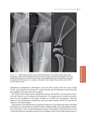
Figure 15.2 Radiographic anatomy of the (normal) shoulder joint: (A) lateral and (B) craniocaudal SHOULDER REGION
radiographic view of a normal adult shoulder; (C) lateral and (D) craniocaudal radiographic view of an
immature dog, wherein the white arrows indicate normal appearance of the proximal humerus physis;
(E) separation of the humerus in a cadaver, to illustrate normal shape of the proximal physis of the
humerus (white arrow).
infraspinatus, supraspinatus, subscapularis, and teres minor muscles. The teres major, biceps
brachii, caput longum of the triceps, the coracobrachialis, and the deltoideus muscles have also
been suggested to provide minor active support.
The tendon of the biceps brachii, subscapularis muscle, and the MGL are located intra-articu-
larly and therefore can be evaluated arthroscopically. The supraspinatus is located extra-articu-
larly. The main stabilizers of the medial shoulder are the MGL and subscapularis, while the
tendons of the supraspinatus, infraspinatus, and teres minor muscles and the LGL provide the
majority of the lateral stability.
Osteoarthritis of the shoulder joint is generally thought to be associated with common shoulder
pathologies (e.g. osteochondrosis, medial shoulder instability [MSI], and tendinopathies) indicat-
ing a secondary (i.e. due to underlying disease) etiology. Interestingly, a study found that over 50%
of dogs necropsied for unrelated reasons showed cartilage erosion of the caudal humeral head, of