
Page 267 - Canine Lameness
P. 267
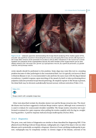
15.6 iceps rachii Tendinopathy 239
(A) (B) (C)
Figure 15.10 Cadaveric specimen demonstrating the biceps brachii anatomy of the medial aspect of the
shoulder: (A) superficial dissection illustrating the structures overlaying the biceps tendon; (B) exposure of
the biceps after removal of the pectoralis musculature; and (C) after transaction of the transverse humeral
ligament and elevation of the supraspinatus muscle, the entire tendon of the biceps brachii can be seen
deeply seated in the intertubercular groove: (a) supraspinatus muscle; (b) superficial pectoralis; (c) greater
tubercle; and (d) biceps brachii.
entire muscle should be performed in this position. Some dogs may resist this test in a standing
position because of other pathologies in the contralateral limb. As it is typically not known if there
is bilateral disease or not, it is recommended to also perform the same exam with the dog in lateral
recumbency. A painful response upon stretching of the muscle by itself or when performing direct
palpation indicates potential biceps brachii pathology. If complete rupture of the biceps is present,
the distal limb may be elevated caudodorsally beyond normal, without restriction (Video 15.1). SHOULDER REGION
Video 15.1
Biceps stretch with complete biceps tear.
Other tests described include the shoulder drawer test and the biceps retraction test. The shoul-
der drawer test has been suggested to indicate biceps tendon rupture, although most commonly it
is used to evaluate for craniocaudal shoulder instability. The biceps tendon retraction test is per-
formed to evaluate for a pain response when pulling the tendon caudally with the dog in a weight-
bearing position. A positive response indicates biceps tendinopathy (Rochat 2018).
15.6.3 Diagnostics
The pros, cons, and choice of diagnostics are similar to those described for diagnosing MSI. If the
physical exam findings indicate biceps disease, radiographs are a logical first step to rule out other
diseases and to potentially accomplish a diagnosis. However, if non-calcifying tendinopathy is pre-
sent, radiographs may be completely normal. In chronic stages of the disease, sclerosis of the