
Page 269 - Canine Lameness
P. 269
15.6 iceps rachii Tendinopathy 241
(A) (B) (C)
(D)
(E) (F)
(G)
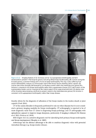
Figure 15.12 Imaging features of (A–D) biceps and (E–G) supraspinatus tendinopathy: (A) faint
mineralization present in the bicipital groove, indicating likely biceps tendinopathy; (B) sclerosis along the
bicipital groove, a common finding with chronic bicipital tendinopathy. This dog is also suffering from
severe osteoarthritic changes; (C) arthroscopic view of the biceps tendon showing partial rupture; (D) SHOULDER REGION
skyline view of the shoulder, demonstrating calcification present within the intertubercular groove, this
location is consistent with biceps tendinopathy rather than supraspinatus disease; (E, F) calcification of the
supraspinatus tendon can be visualized at the cranial aspect of the scapulohumeral joint; (G) skyline view
of the shoulder, demonstrating calcification present lateral to the intertubercular groove, this location is
consistent with supraspinatus tendinopathy rather than biceps disease.
thereby allows for the diagnosis of adhesions of the biceps tendon to the tendon sheath or joint
capsule (Cook 2016).
CT of the canine shoulder is frequently performed to rule out other diseases but it is not consid-
ered a primary imaging modality for biceps tendinopathy. CT arthrography is reported to be of
more diagnostic value than CT alone in diagnosing biceps pathology, and CT arthrography with
epinephrine appears to improve image sharpness, particularly if imaging is delayed (De Simone
et al. 2013; Eivers et al. 2018).
MRI (Figure 10.5) is a sensitive diagnostic tool for identifying both primary biceps tendinopathy
and biceps impingement (Murphy et al. 2008).
Arthroscopy has the distinct advantage to be able to combine diagnostic value with potential
immediate therapy (e.g. biceps tendon release).