
Page 277 - Canine Lameness
P. 277
15.10 atto rOtmOtO ootaarAe attd tNtalnto terNA 249
return of shoulder ROM and function regardless of intervention (Eljabu et al. 2016). This condition
is well described in people and has recently been reported in eight canine patients (Carr et al.
2016). All patients were reported to have a chronic, severe thoracic limb lameness with pain on
shoulder manipulation and severe decrease in ROM. None of the patients responded to treatment.
Diagnostic imaging such as MRI, ultrasound, and arthroscopy may be utilized to identify underly-
ing pathology. A tentative diagnosis is made if excessive joint capsule fibrosis, scar tissue, or adhe-
sion formation is observed (Figure 15.16). If other shoulder conditions are observed, it is difficult
to know whether the observed fibrotic changes are consequences of the other disease processes or
a separate disease process. Successful treatment of adhesive capsulitis has not yet been identified
in dogs. However, the thawing phase in people is reached one to three years after initial onset of
symptoms (Eljabu et al. 2016) and it is unknown when or if this phase occurs in dogs.
15.10.4 Shoulder Region Neoplasia
The most common neoplasia of the shoulder is proximal humeral osteosarcoma. Brachial plexus
neoplasia may also mimic a shoulder lameness. These conditions should be considered as differen-
tial diagnoses in any patient and are described in Chapters 11, 16, and 17.
(A) (C) (E)
(F)
(B) (D) SHOULDER REGION
(G)
(H)
(I)
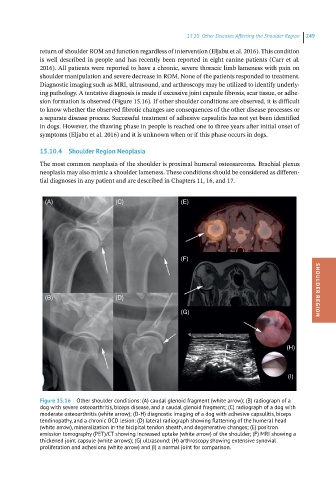
Figure 15.16 Other shoulder conditions: (A) caudal glenoid fragment (white arrow); (B) radiograph of a
dog with severe osteoarthritis, biceps disease, and a caudal glenoid fragment; (C) radiograph of a dog with
moderate osteoarthritis (white arrow); (D-H) diagnostic imaging of a dog with adhesive capsulitis, biceps
tendinopathy, and a chronic OCD lesion: (D) lateral radiograph showing flattening of the humeral head
(white arrow), mineralization in the bicipital tendon sheath, and degenerative changes; (E) positron
emission tomography (PET)/CT showing increased uptake (white arrow) of the shoulder; (F) MRI showing a
thickened joint capsule (white arrows); (G) ultrasound; (H) arthroscopy showing extensive synovial
proliferation and adhesions (white arrow) and (I) a normal joint for comparison.