
Page 340 - Canine Lameness
P. 340
312 19 Stifle Region
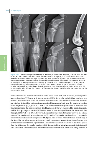
(A) (C) (E) (G)
(B) (D) (F)
STIFLE REGION
Figure 19.2 Normal radiographic anatomy of the stifle joint (Note: for images B, D lateral is on the left):
(A, B, E, F) lateral and craniocaudal views of the stifle of adult dogs; (C, D, G) lateral and craniocaudal
views of the stifle of immature dogs; (a) femur; (b) tibia; (c) patella; (d) fibula; (e) fabella(e), i.e. lateral/
medial gastrocnemius sesamoid bone; (f) popliteal sesamoid bone; (g) proximal tibial physis; (h) distal
femur physis; (i) apophysis of tibial tuberosity; (j) extensor fossa, i.e. fossa of origin of the long digital
extensor tendon; (k) medial and lateral supracondylar tuberosities (i.e. origin of the gastrocnemius
muscle) and superficial digital flexor (lateral tuberosity); (l) tibial tuberosity; (m) femorotibial joint; (n)
femoropatellar joint; (o) patellar ligament; (p) infrapatellar fat pad; and (q) cranial and caudal horn of the
menisci/joint fluid.
meniscal horns and attachments are nerve and blood vessel rich and, therefore, have important
sensory functions (O’Connor and Mcconnaughey 1978). Regionally, an individual meniscus con-
sists of a body and a cranial and caudal horn. The cranial and caudal horns of the medial meniscus
are attached to the tibial plateau via meniscotibial ligaments, which hold the meniscus in place
while weight-bearing (Pagnani et al. 1991). The transverse (formerly described as intermeniscal)
ligament connects the cranial menisco-tibial ligaments of the two menisci. The menisci are quite
mobile through range of motion (ROM) and move to match the position of the femur and tibia
through ROM (Park et al. 2018). However, there are two important differences between the attach-
ments of the medial and the lateral meniscus. The body of the medial meniscus has a close associa-
tion with the medial collateral ligament (MCL) and joint capsule, which tethers it more firmly to
the tibia. The lateral meniscus, on the other hand, has a strong association with the femoral con-
dyle via the menisco-femoral ligament that anchors the caudal meniscal horn to the femur (rather
than the tibia since the caudal menisco-tibial ligament of the lateral meniscus is infinitesimal).
This association allows the lateral meniscus to move with the femur, rather than being tethered to