
Page 397 - Canine Lameness
P. 397
20.7 uscle Contractures 369
affected limb with reduced stifle extension and a quick, almost elastic jerking motion of the limb
with internal/medial rotation of the foot with external rotation of the calcaneus and concurrent
internal rotation of the stifle during the mid and late swing phase (Video 20.4). In the early stages
of the disease, these changes in gait can be subtle. However, the disease typically progresses until
it becomes pathognomonic for gracilis contracture.
Video 20.4:
Gracilis contracture gait.
20.7.1.2 Physical Exam
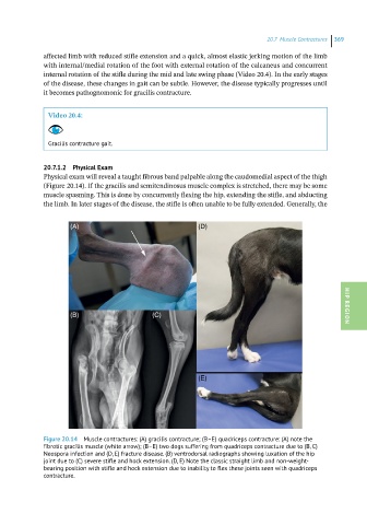
Physical exam will reveal a taught fibrous band palpable along the caudomedial aspect of the thigh
(Figure 20.14). If the gracilis and semitendinosus muscle complex is stretched, there may be some
muscle spasming. This is done by concurrently flexing the hip, extending the stifle, and abducting
the limb. In later stages of the disease, the stifle is often unable to be fully extended. Generally, the
(A) (D)
(B) (C) HIP REGION
(E)
Figure 20.14 Muscle contractures: (A) gracilis contracture; (B–E) quadriceps contracture: (A) note the
fibrotic gracilis muscle (white arrow); (B–E) two dogs suffering from quadriceps contracture due to (B, C)
Neospora infection and (D, E) fracture disease. (B) ventrodorsal radiographs showing luxation of the hip
joint due to (C) severe stifle and hock extension. (D, E) Note the classic straight limb and non-weight-
bearing position with stifle and hock extension due to inability to flex these joints seen with quadriceps
contracture.