
Page 400 - Canine Lameness
P. 400
372 20 Hip Region
(A) (C)
(B) (D)
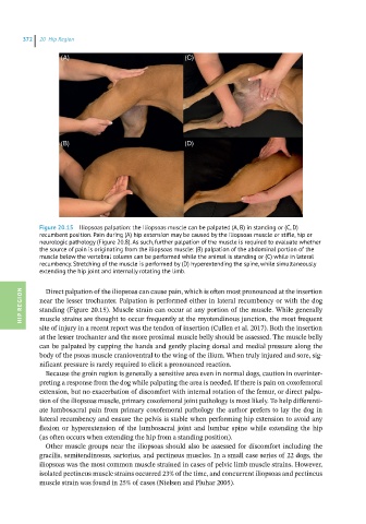
Figure 20.15 Iliopsoas palpation: the Iliopsoas muscle can be palpated (A, B) in standing or (C, D)
recumbent position. Pain during (A) hip extension may be caused by the iliopsoas muscle or stifle, hip or
neurologic pathology (Figure 20.8). As such, further palpation of the muscle is required to evaluate whether
the source of pain is originating from the iliopsoas muscle: (B) palpation of the abdominal portion of the
muscle below the vertebral column can be performed while the animal is standing or (C) while in lateral
recumbency. Stretching of the muscle is performed by (D) hyperextending the spine, while simultaneously
extending the hip joint and internally rotating the limb.
Direct palpation of the iliopsoas can cause pain, which is often most pronounced at the insertion
HIP REGION near the lesser trochanter. Palpation is performed either in lateral recumbency or with the dog
standing (Figure 20.15). Muscle strain can occur at any portion of the muscle. While generally
muscle strains are thought to occur frequently at the myotendinous junction, the most frequent
site of injury in a recent report was the tendon of insertion (Cullen et al. 2017). Both the insertion
at the lesser trochanter and the more proximal muscle belly should be assessed. The muscle belly
can be palpated by cupping the hands and gently placing dorsal and medial pressure along the
body of the psoas muscle cranioventral to the wing of the ilium. When truly injured and sore, sig-
nificant pressure is rarely required to elicit a pronounced reaction.
Because the groin region is generally a sensitive area even in normal dogs, caution in overinter-
preting a response from the dog while palpating the area is needed. If there is pain on coxofemoral
extension, but no exacerbation of discomfort with internal rotation of the femur, or direct palpa-
tion of the iliopsoas muscle, primary coxofemoral joint pathology is most likely. To help differenti-
ate lumbosacral pain from primary coxofemoral pathology the author prefers to lay the dog in
lateral recumbency and ensure the pelvis is stable when performing hip extension to avoid any
flexion or hyperextension of the lumbosacral joint and lumbar spine while extending the hip
(as often occurs when extending the hip from a standing position).
Other muscle groups near the iliopsoas should also be assessed for discomfort including the
gracilis, semitendinosus, sartorius, and pectineus muscles. In a small case series of 22 dogs, the
iliopsoas was the most common muscle strained in cases of pelvic limb muscle strains. However,
isolated pectineus muscle strains occurred 23% of the time, and concurrent iliopsoas and pectineus
muscle strain was found in 25% of cases (Nielsen and Pluhar 2005).