
Page 403 - Canine Lameness
P. 403
20.9 ctrF Hesrresres ooracHoe ctrf Hip reH o 375
20.9.5 Miscellaneous Other Conditions
Sorjonen et al. (1990) described compression of the sciatic nerve between the sacrotuberous l igament
and secondary changes from severe hip OA.
Calcification of the gluteal muscles has been described and may indicate chronic trauma or overuse
(Liu and Dorfman 1976).
MacInnes et al. reported a benign unicameral bone cyst of the hip joint causing mild lameness in
an 11 year old dog (MacInnes et al. 2005).
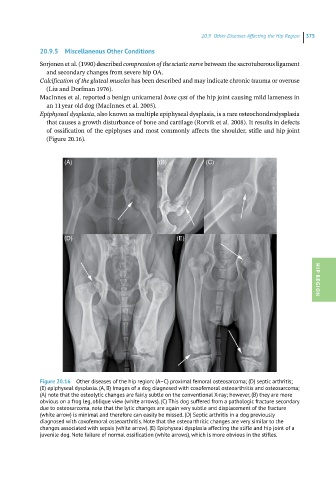
Epiphyseal dysplasia, also known as multiple epiphyseal dysplasia, is a rare osteochondrodysplasia
that causes a growth disturbance of bone and cartilage (Rorvik et al. 2008). It results in defects
of ossification of the epiphyses and most commonly affects the shoulder, stifle and hip joint
(Figure 20.16).
(A) (B) (C)
(D) (E) HIP REGION
Figure 20.16 Other diseases of the hip region: (A–C) proximal femoral osteosarcoma; (D) septic arthritis;
(E) epiphyseal dysplasia. (A, B) Images of a dog diagnosed with coxofemoral osteoarthritis and osteosarcoma;
(A) note that the osteolytic changes are fairly subtle on the conventional X-ray; however, (B) they are more
obvious on a frog leg, oblique view (white arrows). (C) This dog suffered from a pathologic fracture secondary
due to osteosarcoma, note that the lytic changes are again very subtle and displacement of the fracture
(white arrow) is minimal and therefore can easily be missed. (D) Septic arthritis in a dog previously
diagnosed with coxofemoral osteoarthritis. Note that the osteoarthritic changes are very similar to the
changes associated with sepsis (white arrow). (E) Epiphyseal dysplasia affecting the stifle and hip joint of a
juvenile dog. Note failure of normal ossification (white arrows), which is more obvious in the stifles.