
Page 413 - Canine Lameness
P. 413
21.3 Neurorogico giNciNi AANicgio ctNe Nolgi gim 385
amount of sclerosis adjacent to the lytic regions. Because radiographic abnormalities are often
delayed, establishing a diagnosis may require advanced imaging (CT, MRI, or nuclear imaging).
Alternatively, serial radiographs may be appropriate, particularly if positive culture results support
the tentative diagnosis of discospondylitis.
Identification of the infectious agent is important to determine appropriate treatment. Prior to
initiation of antibiotic therapy, urine and blood culture samples should be obtained. A thorough
physical examination is also important to detect other sites of infection, such as bacterial endocar-
ditis, prostatitis, skin, teeth, and ear canals. Combining bacterial cultures of urine and blood is
thought to provide the greatest chance in isolating the causal organism and infection by multiple
agents is reported. Testing for Brucella canis is critical from a zoonotic standpoint. More invasive
techniques have been used such as fluoroscopically guided percutaneous needle aspiration of the
IVD and surgical biopsies with variable success.
(A) (B)
(C) (D)
(E) (F)
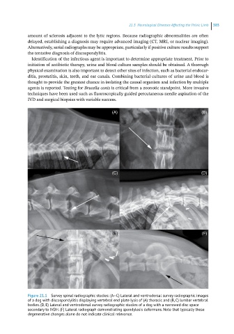
Figure 21.1 Survey spinal radiographic studies: (A–C) Lateral and ventrodorsal survey radiographic images
of a dog with discospondylitis displaying vertebral end plate lysis of (A) thoracic and (B, C) lumbar vertebral
bodies. (D, E) Lateral and ventrodorsal survey radiographic studies of a dog with a narrowed disc space
secondary to IVDH. (F) Lateral radiograph demonstrating spondylosis deformans. Note that typically these
degenerative changes alone do not indicate clinical relevance.