
Page 422 - Canine Lameness
P. 422
394 22 Neoplastic Conditions of the Pelvic Limb
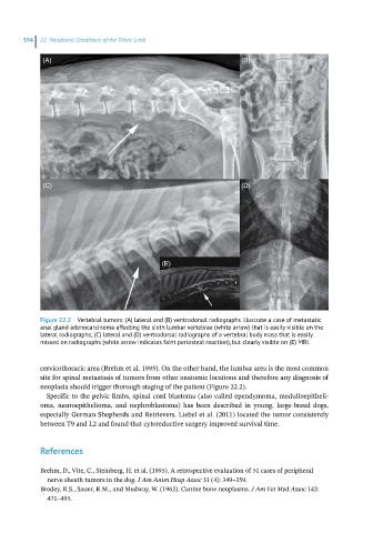
(A) (B)
(C) (D)
(E)
Figure 22.2 Vertebral tumors: (A) lateral and (B) ventrodorsal radiographs illustrate a case of metastatic
anal gland adenocarcinoma affecting the sixth lumbar vertebrae (white arrow) that is easily visible on the
lateral radiographs; (C) lateral and (D) ventrodorsal radiographs of a vertebral body mass that is easily
missed on radiographs (white arrow indicates faint periosteal reaction), but clearly visible on (E) MRI.
cervicothoracic area (Brehm et al. 1995). On the other hand, the lumbar area is the most common
site for spinal metastasis of tumors from other anatomic locations and therefore any diagnosis of
neoplasia should trigger thorough staging of the patient (Figure 22.2).
Specific to the pelvic limbs, spinal cord blastoma (also called ependymoma, medulloepitheli-
oma, neuroepithelioma, and nephroblastoma) has been described in young, large‐breed dogs,
especially German Shepherds and Retrievers. Liebel et al. (2011) located the tumor consistently
between T9 and L2 and found that cytoreductive surgery improved survival time.
References
Brehm, D., Vite, C., Steinberg, H. et al. (1995). A retrospective evaluation of 51 cases of peripheral
nerve sheath tumors in the dog. J Am Anim Hosp Assoc 31 (4): 349–359.
Brodey, R.S., Sauer, R.M., and Medway, W. (1963). Canine bone neoplasms. J Am Vet Med Assoc 143:
471–495.