
Page 45 - Canine Lameness
P. 45
2.2 inetic Analysis 17
Portability and Storage – Most veterinary FP systems are permanently affixed to the ground in a
dedicated space. The limited mobility of these systems should be considered by clinicians with-
out dedicated space or where portability is of importance.
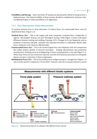
2.2.2 Force Plate Systems: Kinetic Measurements
FP systems measure force in three directions: (i) vertical force, (ii) craniocaudal force, and (iii)
mediolateral force (Figure 2.2):
Vertical Force (Fz) – This is the largest and most commonly evaluated force. Graphically, it
appears “bell‐shaped” during a trot and “M‐shaped” during a walk (Figure 2.3) due to the speed
difference between trotting and walking (Decamp 1997). Changes in Fz are frequently used as
measures of function and pain. Animals with lameness secondary to orthopedic disease often
have a reduction in Fz values in affected limbs.
Craniocaudal Force (Fy) – This is the second largest force and is biphasic with two components
that define distinct periods of forward movement – braking (deceleration) and propulsion
(acceleration). Braking occurs at the beginning of stance and propulsion at the end (Figure 2.3).
While these forces are less commonly reported, animals with lameness secondary to orthopedic
disease can have a reduction in these components.
Mediolateral Force (Fx) – This is the smallest force in dogs moving in a straight line (Figure 2.3)
and is rarely used for comparison (Torres 2018). However, with the increased interest in canine
Figure 2.2 Measurements that can be obtained with different kinetic systems.