
Page 50 - Canine Lameness
P. 50
22 2 Objective Gait Analysis
In veterinary medicine, 2D analysis is most common. While 3D analysis provides a more complete
representation of actual movement, it is complex to perform and requires advanced equipment.
This consideration needs to be accounted for when deciding what is most appropriate in a particu-
lar clinical setting.
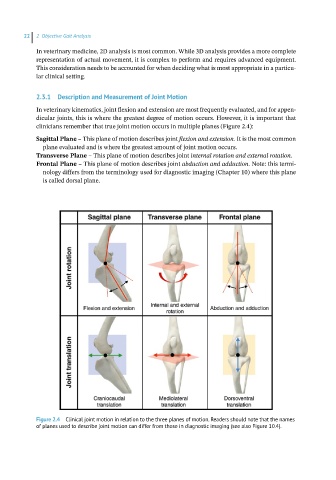
2.3.1 Description and Measurement of Joint Motion
In veterinary kinematics, joint flexion and extension are most frequently evaluated, and for appen-
dicular joints, this is where the greatest degree of motion occurs. However, it is important that
clinicians remember that true joint motion occurs in multiple planes (Figure 2.4):
Sagittal Plane – This plane of motion describes joint flexion and extension. It is the most common
plane evaluated and is where the greatest amount of joint motion occurs.
Transverse Plane – This plane of motion describes joint internal rotation and external rotation.
Frontal Plane – This plane of motion describes joint abduction and adduction. Note: this termi-
nology differs from the terminology used for diagnostic imaging (Chapter 10) where this plane
is called dorsal plane.
Figure 2.4 Clinical joint motion in relation to the three planes of motion. Readers should note that the names
of planes used to describe joint motion can differ from those in diagnostic imaging (see also Figure 10.4).