
Page 51 - Canine Lameness
P. 51
2.3 inematic Analysis 23
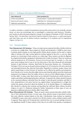
Box 2.1 Six Degrees of Freedom (6‐DOF) Movement
Joint rotations (3) Joint translations (3)
Flexion and extension Craniocaudal or “craniocaudal translation”
Internal and external rotation Mediolateral or “mediolateral translation”
Abduction and adduction Dorsoventral or “joint distraction”
In order to translate a complex biomechanical process like joint motion into clinically relevant
terms, we must use terminology that is meaningful to researchers and clinicians. Therefore,
joint motion is often discussed using the concept of six degrees of freedom (6‐DOF; Grood and
Suntay 1983). This describes clinical joint motion relative to three axes (x, y, and z) and associ-
ated with these axes are six distinct motions consisting of (3) rotations and (3) translations
(Figure 2.4; Box 2.1).
2.3.2 Kinematic Systems
Two‐Dimensional (2D) Systems – These are single camera systems that allow reliable collection
of motion in a single plane. These systems are simple and inexpensive (<$1000 in most cases).
Consumer grade cameras or even high‐quality cellular phone cameras can be used to capture
video of animals with high contrast spherical markers applied to the skin or coat (e.g. white
markers on a black dog or black markers on a white dog). These markers can then be digitized
by computerized software programs to determine joint or body movement. There are numerous
software programs for 2D kinematics. Kinovea (www.kinovea.org), for example, is a free and
open source program that is easy to use but lacks some of the more advanced and automated
features of paid software. Likewise, MaxTRAQ 2D (Innovision Systems, Inc., Columbiavile,
Michigan, USA) is an easy‐to‐use paid software program for 2D kinematics with many advanced
and automated features that clinicians find useful. Regardless of software, proper camera and
patient positioning is critical because 2D systems can be affected by parallax and perspective
error. Parallax error occurs when the subject moves away from the optical axis of the camera and
perspective error happens when the subject moves in and out of the calibrated plane of motion
(Kirtley 2006). In many cases, these errors can be reduced if clinicians pay careful attention to
camera and subject positioning during data collection. In general, the camera should be placed
at the maximum distance allowable from the subject to provide good resolution and field of
view, perpendicular to the plane interest (e.g. sagittal plane), and positioned at a height that
centers the camera at the level where the markers are to be tracked (Figure 2.5). Additionally,
data should be collected from patients moving along the calibrated plane without deviation
(Figures 2.6 and 2.7). Clinicians wishing for further information on these types of error are
directed to additional resources (Kirtley 2006; Torres 2018).
Three‐Dimensional (3D) Systems – These are multi‐camera systems that allow reliable collec-
tion of three‐dimensional motion. In most cases, these systems consist of specialized cameras
that track reflective markers placed on the skin. Unfortunately, these systems are expensive
(>$100 000 in many cases) and to evaluate true 3D joint motion a more complex marker model
is required. These aspects of 3D systems have restricted their use to research settings. However,
one significant benefit of these systems is that they do not suffer from parallax or perspective