
Page 60 - Canine Lameness
P. 60
32 3 The Orthopedic Examination
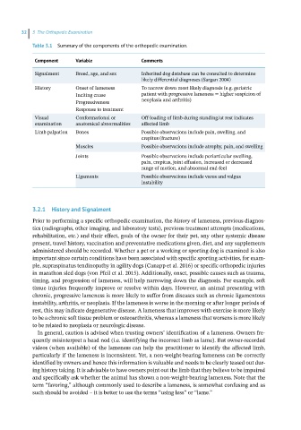
Table 3.1 Summary of the components of the orthopedic examination.
Component Variable Comments
Signalment Breed, age, and sex Inherited dog database can be consulted to determine
likely differential diagnoses (Sargan 2004)
History Onset of lameness To narrow down most likely diagnosis (e.g. geriatric
Inciting cause patient with progressive lameness = higher suspicion of
Progressiveness neoplasia and arthritis)
Response to treatment
Visual Conformational or Off‐loading of limb during standing/at rest indicates
examination anatomical abnormalities affected limb
Limb palpation Bones Possible observations include pain, swelling, and
crepitus (fracture)
Muscles Possible observations include atrophy, pain, and swelling
Joints Possible observations include periarticular swelling,
pain, crepitus, joint effusion, increased or decreased
range of motion, and abnormal end‐feel
Ligaments Possible observations include varus and valgus
instability
3.2.1 History and Signalment
Prior to performing a specific orthopedic examination, the history of lameness, previous diagnos-
tics (radiographs, other imaging, and laboratory tests), previous treatment attempts (medications,
rehabilitation, etc.) and their effect, goals of the owner for their pet, any other systemic disease
present, travel history, vaccination and preventative medications given, diet, and any supplements
administered should be recorded. Whether a pet or a working or sporting dog is examined is also
important since certain conditions have been associated with specific sporting activities, for exam-
ple, supraspinatus tendinopathy in agility dogs (Canapp et al. 2016) or specific orthopedic injuries
in marathon sled dogs (von Pfeil et al. 2015). Additionally, onset, possible causes such as trauma,
timing, and progression of lameness, will help narrowing down the diagnosis. For example, soft
tissue injuries frequently improve or resolve within days. However, an animal presenting with
chronic, progressive lameness is more likely to suffer from diseases such as chronic ligamentous
instability, arthritis, or neoplasia. If the lameness is worse in the morning or after longer periods of
rest, this may indicate degenerative disease. A lameness that improves with exercise is more likely
to be a chronic soft tissue problem or osteoarthritis, whereas a lameness that worsens is more likely
to be related to neoplasia or neurologic disease.
In general, caution is advised when trusting owners’ identification of a lameness. Owners fre-
quently misinterpret a head nod (i.e. identifying the incorrect limb as lame). But owner‐recorded
videos (when available) of the lameness can help the practitioner to identify the affected limb,
particularly if the lameness is inconsistent. Yet, a non‐weight‐bearing lameness can be correctly
identified by owners and hence this information is valuable and needs to be clearly teased out dur-
ing history taking. It is advisable to have owners point out the limb that they believe to be impaired
and specifically ask whether the animal has shown a non‐weight‐bearing lameness. Note that the
term “favoring,” although commonly used to describe a lameness, is somewhat confusing and as
such should be avoided – it is better to use the terms “using less” or “lame.”