
Page 64 - Canine Lameness
P. 64
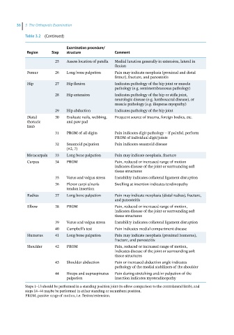
36  3  The Orthopedic Examination

            Table 3.2  (Continued)

                              Examination procedure/
             Region    Step   structure            Comment

                         25   Assess location of patella  Medial luxation generally in extension, lateral in
                                                   flexion
             Femur       26   Long bone palpation  Pain may indicate neoplasia (proximal and distal
                                                   femur), fracture, and panosteitis
             Hip         27   Hip flexion          Indicates pathology of the hip joint or muscle
                                                   pathology (e.g. semimembranosus pathology)
                         28   Hip extension        Indicates pathology of the hip or stifle joint,
                                                   neurologic disease (e.g. lumbosacral disease), or
                                                   muscle pathology (e.g. iliopsoas myopathy)
                         29   Hip abduction        Indicates pathology of the hip joint
             Distal      30   Evaluate nails, webbing,   Frequent source of trauma, foreign bodies, etc.
             thoracic         and paw pad
             limb
                         31   PROM of all digits   Pain indicates digit pathology – if painful, perform
                                                   PROM of individual digit/joints
                         32   Sesamoid palpation    Pain indicates sesamoid disease
                              (#2, 7)
             Metacarpals  33  Long bone palpation  Pain may indicate neoplasia, fracture
             Carpus      34   PROM                 Pain, reduced or increased range of motion
                                                   indicates disease of the joint or surrounding soft
                                                   tissue structures
                         35   Varus and valgus stress  Instability indicates collateral ligament disruption
                         36   Flexor carpi ulnaris   Swelling at insertion indicates tendinopathy
                              tendon insertion
             Radius      37   Long bone palpation  Pain may indicate neoplasia (distal radius), fracture,
                                                   and panosteitis
             Elbow       38   PROM                 Pain, reduced or increased range of motion,
                                                   indicates disease of the joint or surrounding soft
                                                   tissue structures
                         39   Varus and valgus stress  Instability indicates collateral ligament disruption
                         40   Campbell’s test      Pain indicates medial compartment disease
             Humerus     41   Long bone palpation  Pain may indicate neoplasia (proximal humerus),
                                                   fracture, and panosteitis
             Shoulder    42   PROM                 Pain, reduced or increased range of motion,
                                                   indicates disease of the joint or surrounding soft
                                                   tissue structures
                         43   Shoulder abduction   Pain or increased abduction angle indicates
                                                   pathology of the medial stabilizers of the shoulder
                         44   Biceps and supraspinatus   Pain during stretching and/or palpation of the
                              palpation            insertion indicates myotendinopathy
            Steps 1–13 should be performed in a standing position joint (to allow comparison to the contralateral limb), and
            steps 14–44 maybe be performed in either standing or recumbent position.
            PROM, passive range of motion, i.e. flexion/extension.