
Page 504 - Clinical Small Animal Internal Medicine
P. 504
472 Section 6 Gastrointestinal Disease
bodies and obstructive intestinal disease in small ani Commonly used neutral attenuation luminal contrast
VetBooks.ir mals has not been shown to have statistically significant agents include water, milk, polyethylene glycol, 12.5%
corn‐oil emulsion, and methylcellulose.
increased sensitivity or specificity compared to plain
radiography. In gastrointestinal CT without the use of
intraluminal contrast agents, the lack of distension of the Magnetic Resonance Imaging
small intestine hinders evaluation of the intestinal wall of the Gastrointestinal Tract
throughout the entire gastrointestinal tract.
In human medicine, it has been well demonstrated Magnetic resonance imaging is rarely used for evaluation
that a critical technical requirement for CT of the bowel of the gastrointestinal tract in human and veterinary
is full distension of a clear lumen with complete separa patients. In human patients, new MRI applications for
tion of the intestinal walls. In CT enterography, combi imaging of the colon are being developed. The most
nations of positive and neutral attenuation oral contrast common use of MRI related to digestive disease in vet
agents are used, which allow distension of the intestinal erinary patients involves imaging of the oral cavity or
tract and outlining of the wall. Conventional positive cranial neck in patients where abscesses secondary to
attenuation oral contrast media such as barium‐ or perforating injuries or foreign bodies are suspected.
iodine‐based agents are used for evaluation of the gas
trointestinal tract similarly to their use in radiography or Gastrointestinal Scintigraphy
fluoroscopy (Figure 48.2). Conventional positive attenu
ation oral contrast agents allow visualization of the Ultrasound and contrast radiography continue to be the
mucosal detail and assessment of mucosal enhancement. most common imaging techniques used for the gastroin
Neutral oral contrast agents, which are more commonly testinal tract. However, contrast radiography, similarly to
used in human medicine, have an attenuation close to contrast‐enhanced CT, provides only very limited or no
water and are mainly used to distend the small intesti information about esophageal or gastrointestinal motil
nal tract and therefore better outline the intestinal ity. It has been suggested that esophageal scintigraphy
walls, allowing for better evaluation for mural lesions. might provide valuable information in gastroesophageal
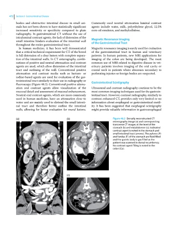
(a) (b) Figure 48.2 Dorsally reconstructed CT
enterography image (a) and corresponding
transverse CT images at the level of the
stomach (b) and midabdomen (c). Iodinated
contrast agent is noted in the stomach and
small intestinal tract (arrows). The pylorus (P)
and fundus (F) of the stomach are fluid filled
and the gastric body is gas filled as the
patient was scanned in dorsal recumbency.
No contrast agent filling is noted in the
colon (Co).
(c)