
Page 507 - Clinical Small Animal Internal Medicine
P. 507
48 Gastrointestinal Imaging 475
of the esophagus results, barium paste should be used to It has been noted in studies in humans as well as experi
VetBooks.ir enhance coating of the mucosa. mentally in rats that no significant mediastinitis was
identified post barium leakage through a very small
Double contrast esophagus studies are commonly per
formed in human patients but are rarely used in veteri
with subsequent radiographic evaluations noted. It has
nary medicine. In some patients, where air is present esophageal defect, nor was a significant interference
in the dilated esophagus, it functions as an endogenous been recommended to warm up the barium solution to
negative contrast agent and no additional contrast 40 °C when a perforation is suspected, as this decreases
medium may be needed. If barium sulfate suspension is the viscosity of the barium sulfate solution without
used for the esophagram, no contrast should be remain decreasing the radiopacity and increases the sensitivity
ing in the esophagus in approximately 5 minutes; if bar of detecting smaller sites of leakage in the esophagus.
ium paste is used, all contrast agent should be cleared
from the esophagus by approximately 30 minutes. In Generalized Esophageal Dilation (Megaesophagus)
general, in the normal esophagus, no contrast agent
should be stored or remain after a contrast agent study Megaesophagus is a disorder caused by diminished
has been performed. esophageal tone and hypomotility, causing generalized
In patients with suspected esophageal perforation, dilation of the esophagus. Congenital and acquired forms
water‐soluble contrast solutions should be used for eval of megaesophagus are recognized in the dog and cat. The
uation of potential leakage from the esophagus. However, underlying cause of congenital megaesophagus remains
water‐soluble iodine solution contrast agents should be unknown, but inadequate development of the esopha
used with great care for an esophagram, as ionic iodi geal innervation or altered biochemical properties of the
nated contrast agents are hypertonic and, when aspi esophagus have been discussed.
rated, will most likely cause severe pulmonary edema. If Survey radiographs are often diagnostic for a dilated
a bronchoesophageal fistula is suspected, nonionic iodi esophagus, when the esophagus contains food, water or
nated contrast solutions should be used to reduce the air (Figure 48.3).
risk of iatrogenic pulmonary edema. Water‐soluble con On lateral radiographs, the dilated esophagus is usually
trast agents for outlining the esophagus are usually well demarcated dorsally by the longus colli muscle or
administered at a dosage of 2–3 mL/kg bodyweight to summating within the thoracic vertebrae and ventrally
achieve approximately 700 mg iodine per kg bodyweight summating with the dorsal aspect of the trachea, leaving
and adequate distension of the esophagus. However, if the impression of a dorsally thickened trachea wall (the
no contrast leakage is identified, a repeat study with a tracheal stripe sign; Figure 48.4). It is important not to
higher density barium suspension can be performed to confuse the tracheal stripe sign with pneumomediasti
allow visualization of a smaller sized lesion that was num, where the esophagus is circumferentially sur
potentially missed using a water‐soluble contrast agent. rounded by air. In severe esophageal dilation, a ventral
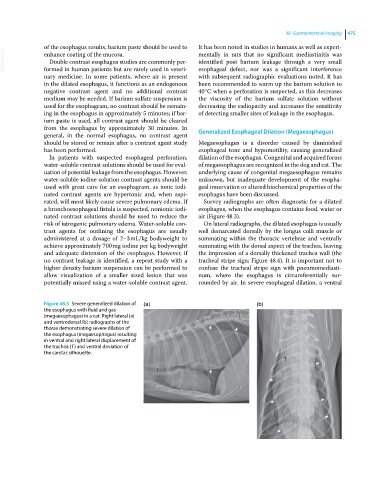
Figure 48.3 Severe generalized dilation of (a) (b)
the esophagus with fluid and gas
(megaesophagus) in a cat. Right lateral (a)
and ventrodorsal (b) radiographs of the
thorax demonstrating severe dilation of
the esophagus (megaesophagus) resulting
in ventral and right lateral displacement of
the trachea (T) and ventral deviation of
the cardiac silhouette.