
Page 510 - Clinical Small Animal Internal Medicine
P. 510
478 Section 6 Gastrointestinal Disease
and cats. The disease is occasionally reported in humans,
VetBooks.ir especially children. The most commonly reported clini
cal signs include spontaneous coughing, coughing after
drinking water, and recurrent chronic respiratory disease.
The most commonly reported fistulae are congenital,
but acquired fistulae secondary to injury of the esopha
gus, such as secondary to a foreign body, have been
observed. The most commonly reported acquired fistulae
are esophagobronchial, with less common reports of
esopha gotracheal and rarely esophagoaortic fistulae. As
the clinical presentation of esophageal fistulae is similar
to patients with aspiration pneumonia, it is crucially
important to differentiate between the two conditions.
It is possible that the fistula opening can be identified
during bronchoscopy or endoscopy of the esophagus;
however, contrast agent swallowing studies using fluor
oscopy are often more reliable for identification of the
connection between the trachea, bronchus or lung and
the esophagus. All images should be carefully evaluated
for leakage of contrast agent outside the esophagus. If a
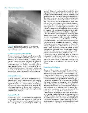
Figure 48.7 Esophageal foreign body. In the cranial cervical fistula is suspected and not noted on an initial esopha
esophagus on a lateral radiograph of the neck of a dog a bone gram, it might be necessary to dilute the barium contrast
surrounded by gas is noted. medium (1:1) as dilution of the barium sulfate suspen
sion decreases the viscosity and therefore allows visuali
Esophagitis, Gastroesophageal Reflux zation of smaller defects in the esophageal wall.
Additional imaging techniques, such as contrast‐
Common causes for esophagitis include gastroesopha enhanced CT, might help to further define the precise
geal reflux, structural abnormalities of the hiatus of the location and size of a fistula. In humans, the use of air as
esophagus (hiatal hernia), neoplasia, trauma, medica a negative contrast agent to outline the esophagus has
tion, and chronic vomiting. Esophagitis is difficult to further helped to demonstrate the presence of the
diagnose on survey radiographs. On an esophagram, a fistula.
caudally thickened and irregular esophagus might be
noted. Radiographically, reflux esophagitis can be con
firmed by filling the stomach with barium and pressing Tumors of the Esophagus
on the stomach. If reflux esophagitis is present, reflux of Tumors of the esophagus are rare, likely accounting for
contrast agent into the esophagus is likely to be noted. less than 0.5% of the tumors reported in cancers in dogs.
Similar epidemiologic studies in cats are currently lacking.
Esophageal Strictures Clinical signs of esophagus disease such as regurgitation
are often only observed when the tumor has grown to
Esophageal strictures can occur secondary to severe ero such a size that it is causing obstruction of the esophagus.
sive esophagitis, and are often associated with perianes In the dog, leiomyomas or leiomyosarcomas affecting
thetic esophageal reflux or secondary to foreign body the lower esophageal sphincter have been described
impaction, trauma or a surgery, where the esophagus (Figure 48.8). Aggressive neoplasms of the esophagus are
was displaced and/or manipulated, such as during ven extremely rare in dogs in areas not endemic for Spirocerca
tral cervical slot surgery. Plain contrast esophagram is lupi. Extremely rarely, squamous cell carcinomas, neu
usually diagnostic, revealing a segment of esophagus that roendocrine carcinoma or adenocarcinomas of the
persistently fails to dilate. esophagus have been reported in the dog and cat.
Endoscopy of the esophagus may allow identification
Esophageal Fistula of esophageal masses. Similarly, contrast radiography
will allow outlining of smaller masses. If the mass is large,
An esophageal fistula is an abnormal connection between it is likely to be noted on survey radiographs (see
the esophagus and trachea, bronchus, lung parenchyma Figure 48.8). Additionally, tumors in the caudal esopha
or skin. Veterinary reports of esophageal fistulae are gus might also be noted during an abdominal ultrasound
rare. Esophageal fistulae are very rarely reported in dogs exam, when the cardia of the stomach is evaluated.