
Page 515 - Clinical Small Animal Internal Medicine
P. 515
48 Gastrointestinal Imaging 483
barium sulfate suspension is in the gastric fundus, while before 20 mL/kg air is added. Four radiographic views of
VetBooks.ir air fills the body and pyloric area of the stomach. Borders the abdomen should be obtained. It is often helpful to
gently roll the patient to achieve an even coating of the
of the gastric wall to luminal contrast agent filling are
normally distinct and well defined.
The diameter of the pylorus is usually approximately gastric wall before the four views are obtained.
half that of the body of the stomach; if it is larger than Gastric Dilation
that, disease of the pyloric canal may be present. The
peristaltic wave normally runs from the fundus to In gastric dilation, the stomach is distended without
the pylorus. During peristalsis, symmetric narrowing of change in shape and position. This can be acute, secondary
the gastric fundus to pylorus wall may be noted. to aerophagia, gastritis, outflow obstruction, medical
Expected times for gastric emptying of contrast are treatment, or chronic, secondary to a chronic obstruc
quite variable. After administration of liquid barium tive process of the pylorus or proximal duodenum.
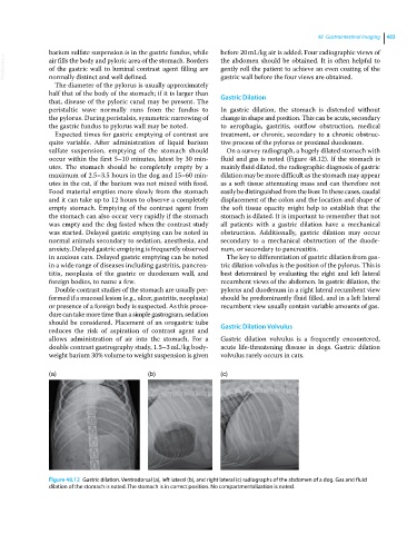
sulfate suspension, emptying of the stomach should On a survey radiograph, a hugely dilated stomach with
occur within the first 5–10 minutes, latest by 30 min fluid and gas is noted (Figure 48.12). If the stomach is
utes. The stomach should be completely empty by a mainly fluid dilated, the radiographic diagnosis of gastric
maximum of 2.5–3.5 hours in the dog and 15–60 min dilation may be more difficult as the stomach may appear
utes in the cat, if the barium was not mixed with food. as a soft tissue attenuating mass and can therefore not
Food material empties more slowly from the stomach easily be distinguished from the liver. In these cases, caudal
and it can take up to 12 hours to observe a completely displacement of the colon and the location and shape of
empty stomach. Emptying of the contrast agent from the soft tissue opacity might help to establish that the
the stomach can also occur very rapidly if the stomach stomach is dilated. It is important to remember that not
was empty and the dog fasted when the contrast study all patients with a gastric dilation have a mechanical
was started. Delayed gastric emptying can be noted in obstruction. Additionally, gastric dilation may occur
normal animals secondary to sedation, anesthesia, and secondary to a mechanical obstruction of the duode
anxiety. Delayed gastric emptying is frequently observed num, or secondary to pancreatitis.
in anxious cats. Delayed gastric emptying can be noted The key to differentiation of gastric dilation from gas
in a wide range of diseases including gastritis, pancrea tric dilation volvulus is the position of the pylorus. This is
titis, neoplasia of the gastric or duodenum wall, and best determined by evaluating the right and left lateral
foreign bodies, to name a few. recumbent views of the abdomen. In gastric dilation, the
Double contrast studies of the stomach are usually per pylorus and duodenum in a right lateral recumbent view
formed if a mucosal lesion (e.g., ulcer, gastritis, neoplasia) should be predominantly fluid filled, and in a left lateral
or presence of a foreign body is suspected. As this proce recumbent view usually contain variable amounts of gas.
dure can take more time than a simple gastrogram, sedation
should be considered. Placement of an orogastric tube Gastric Dilation Volvulus
reduces the risk of aspiration of contrast agent and
allows administration of air into the stomach. For a Gastric dilation volvulus is a frequently encountered,
double contrast gastrography study, 1.5–3 mL/kg body acute life‐threatening disease in dogs. Gastric dilation
weight barium 30% volume to weight suspension is given volvulus rarely occurs in cats.
(a) (b) (c)
Figure 48.12 Gastric dilation. Ventrodorsal (a), left lateral (b), and right lateral (c) radiographs of the abdomen of a dog. Gas and fluid
dilation of the stomach is noted. The stomach is in correct position. No compartmentalization is noted.