
Page 520 - Clinical Small Animal Internal Medicine
P. 520
488 Section 6 Gastrointestinal Disease
(a) (b)
VetBooks.ir
(c)
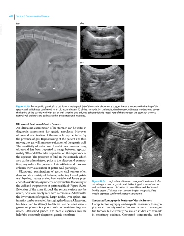
Figure 48.19 Eosinophilic gastritis in a cat. Lateral radiograph (a) of the cranial abdomen is suggestive of a moderate thickening of the
gastric wall, which was confirmed on an ultrasound exam (b) of the stomach. On the longitudinal ultrasound image, moderate to severe
thickening of the gastric wall with loss of wall layering and reduced echogenicity is noted. Part of the fundus of the stomach shows a
normal wall architecture as illustrated in the ultrasound image (c).
Ultrasound Features of Gastric Tumors
An ultrasound examination of the stomach can be useful in
diagnostic assessment for gastric neoplasia. However,
ultrasound examination of the stomach may be limited by
the presence of gas. Repositioning of the patient and thus
moving the gas will improve evaluation of the gastric wall.
The sensitivity of detection of gastric wall masses using
ultrasound has been reported to range between approxi
mately 50% and 80% and is dependent on the experience of
the operator. The presence of fluid in the stomach, which
also can be administered prior to the ultrasound examina
tion, may reduce the presence of air artifacts and therefore
enhance the visualization of gastric wall pathology.
Ultrasound examinations of gastric wall tumors often
demonstrate a variety of features, including loss of gastric
wall layering, masses arising from certain wall layers, pres
ence of cavitations, asymmetric or symmetric thickening of Figure 48.20 Longitudinal ultrasound image of the stomach of a
the wall, and the presence of peritoneal fluid (Figure 48.20). cat. A large, eccentric gastric wall thickening with loss of normal
Extension of the mass through the serosal surface may be wall architecture and distortion of the wall is noted. Peritoneal
fluid is present. This was most concerning for neoplasia. Fine
noted, most commonly seen with carcinoma. Additionally, needle aspirates confirmed a gastric carcinoma.
the involvement of regional lymph nodes, liver, spleen, and
intestine can be evaluated for staging the disease. Ultrasound Computed Tomographic Features of Gastric Tumors
has been used to attempt to differentiate between various Computed tomography and magnetic resonance tomogra
gastric neoplasms, but poor correlation with histology was phy are commonly used in human patients to stage gas
noted. Ultrasound‐guided fine needle aspirates may be tric tumors, but currently no similar studies are available
helpful to accurately diagnose a gastric neoplasm. in veterinary patients. Computed tomography can be