
Page 525 - Clinical Small Animal Internal Medicine
P. 525
48 Gastrointestinal Imaging 493
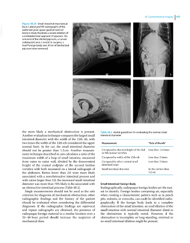
Figure 48.24 Small intestinal mechanical
VetBooks.ir abdomen post upper gastrointestinal
ileus. Lateral and VD radiographs of the
barium study illustrate a severe dilation of
a midabdominal segment of jejunum. On
one end of the dilated jejunum, a curved
radiolucent area is noted. In surgery, a
hoof foreign body and 30 cm of devitalized
jejunum were removed.
the more likely a mechanical obstruction is present. Table 48.2 Useful guidelines for evaluating the normal small
Another evaluation technique compares the largest small intestinal diameter
intestinal diameter with the width of the 12th rib, with
two times the width of the 12th rib considered the upper Measurement “Rule of thumb”
normal limit. In the cat, the small intestinal diameter
should not be greater than 1.2 cm. Another measure Compared to shortest height of the 2nd Less than 1.6 times
ment technique described in cats calculates a ratio of the or 5th lumbar vertebra
maximum width of a loop of small intestine, measured Compared to width of the 12th rib Less than 2 times
from outer to outer wall, divided by the dorsoventral Compared to other normal small Less than 2 times
height of the cranial endplate of the second lumbar intestinal loops
vertebra with both measured on a lateral radiograph of Small intestinal diameter In the cat less than
the abdomen. Ratios lower than 2.0 were more likely 1.2 cm
associated with a nonobstructive intestinal process and
with ratios larger than 3.0, the increased small intestinal
diameter was more than 70% likely to be associated with Small Intestinal Foreign Body
an obstructive intestinal process (Table 48.2). Radiographically, radiopaque foreign bodies are the easi
Single measurements should not be used as the sole est to identify. Foreign bodies containing air, especially
criterion for diagnosis of mechanical obstruction; other when creating a characteristic pattern such as in peach
radiographic findings and the history of the patient pits, walnuts, or corncobs, can easily be identified radio
should be evaluated when considering the differential graphically. If the foreign body leads to a complete
diagnoses. If the radiographic findings are equivocal obstruction of the small intestine, an orad dilation of the
and repeat radiographs are obtained, the presence of small intestine with normal intestinal diameter distal to
radiopaque foreign material in a similar location over a the obstruction is typically noted. However, if the
24–48‐hour period should increase the suspicion of obstruction is incomplete or long‐standing, minimal or
mechanical ileus. no small intestinal dilation might be present.