
Page 526 - Clinical Small Animal Internal Medicine
P. 526
494 Section 6 Gastrointestinal Disease
In an ultrasound examination of the small intestine, intestine and the presence of intestinal intraluminal gas with
VetBooks.ir foreign material in the intestinal tract often appears an abnormal angular or tear drop shape (Figure 48.25).
Perforation of the small intestine can occur, and in addition
hyperechoic with strong distal complete acoustic shad
owing. These shadowing structures often do not move
and free peritoneal gas may be seen (Figure 48.26).
with peristalsis and fluid distension of the small intestine to bunching of the small intestines, a reduced serosal detail
orad to the lesion is often observed. Foreign bodies may Sonographically, an abnormal tortuous path of the
have a distinct shape and surface, which allows differen small intestine, often the duodenum, with a central hyper
tiation of them from food material. echoic, linear structure might be noted. Additionally, an
object with strong distal shadowing might be seen in the
Linear Foreign Body pylorus of the stomach.
Linear foreign bodies cause plication of the small intes
tine and are usually the result of swallowing a linear
object and fixation of part of the foreign material orally Intussusception
or in the stomach. In the cat, linear foreign bodies are Intestinal intussusception can be gastroduondenal,
often fixated around the caudal aspect of the tongue. jejunojejunal (Figure 48.27), ileocolic, cecocolic, and
In the dog, most linear foreign bodies are restrained by colonocolic.
material in the stomach. Radiographically, severely dilated loops of intestine
Classic radiographic features of linear foreign body might be noted. Sometimes a soft tissue attenuation
obstruction include plication or “bunching” of the small mass with orad gas in a convex shape is seen.
Figure 48.25 Linear foreign body.
Ventrodorsal and lateral radiographs of the
abdomen of a cat illustrating multiple
bunched‐up loops of small intestine. On
cross‐section, the luminal gas of the
bunched‐up small intestinal loops has an
abnormal tear drop shape. Both findings
are classic for linear foreign body.
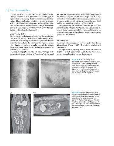
(a) (b) Figure 48.26 Linear foreign body with
perforation. Ventrodorsal (a) and lateral (b)
radiographs of the abdomen post upper
gastrointestinal barium contrast study
demonstrate bunched‐up loops of small
intestine, some with a tear drop shape of
the luminal gas. The reduced serosal detail
and peritoneal gas (arrows) are consistent
with perforation.