
Page 524 - Clinical Small Animal Internal Medicine
P. 524
492 Section 6 Gastrointestinal Disease
Table 48.1 Common imaging findings assessed using ultrasound, Small Intestinal Obstruction (Mechanical Ileus)
VetBooks.ir malignant intestinal wall thickening Radiographic findings in small intestinal obstruction
contrast radiography or computed tomography in benign versus
are dependent on the extent (incomplete or complete),
Benign Malignant location, and duration of obstruction. Small intestinal
obstruction can occur secondary to foreign material,
Symmetric Asymmetric intestinal neoplasia or a mass causing compression of the
Circumferential Eccentric intestinal lumen. Mineral or metal attenuating foreign
Mild to moderate Moderate to severe bodies are radiographically easily detected but non
mineralized and nonmetallic foreign bodies within the
Usually segmental or diffuse Usually focal
intestinal lumen or masses of the small intestinal tract
Frequently abrupt transition
can be challenging to detect. Care should also be taken
Usually normal lumen width Often focally narrowed that mineralizations in the mesenteric fat (mesenteric fat
intestinal lumen
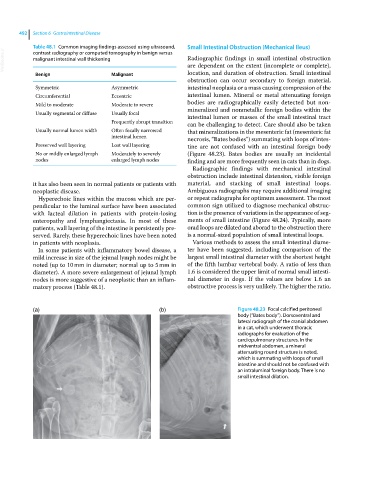
necrosis, “Bates bodies”) summating with loops of intes
Preserved wall layering Lost wall layering tine are not confused with an intestinal foreign body
No or mildly enlarged lymph Moderately to severely (Figure 48.23). Bates bodies are usually an incidental
nodes enlarged lymph nodes finding and are more frequently seen in cats than in dogs.
Radiographic findings with mechanical intestinal
obstruction include intestinal distension, visible foreign
it has also been seen in normal patients or patients with material, and stacking of small intestinal loops.
neoplastic disease. Ambiguous radiographs may require additional imaging
Hyperechoic lines within the mucosa which are per or repeat radiographs for optimum assessment. The most
pendicular to the luminal surface have been associated common sign utilized to diagnose mechanical obstruc
with lacteal dilation in patients with protein‐losing tion is the presence of variations in the appearance of seg
enteropathy and lymphangiectasia. In most of these ments of small intestine (Figure 48.24). Typically, more
patients, wall layering of the intestine is persistently pre orad loops are dilated and aborad to the obstruction there
served. Rarely, these hyperechoic lines have been noted is a normal‐sized population of small intestinal loops.
in patients with neoplasia. Various methods to assess the small intestinal diame
In some patients with inflammatory bowel disease, a ter have been suggested, including comparison of the
mild increase in size of the jejunal lymph nodes might be largest small intestinal diameter with the shortest height
noted (up to 10 mm in diameter; normal up to 5 mm in of the fifth lumbar vertebral body. A ratio of less than
diameter). A more severe enlargement of jejunal lymph 1.6 is considered the upper limit of normal small intesti
nodes is more suggestive of a neoplastic than an inflam nal diameter in dogs. If the values are below 1.6 an
matory process (Table 48.1). obstructive process is very unlikely. The higher the ratio,
(a) (b) Figure 48.23 Focal calcified peritoneal
body (“Bates body”). Dorsoventral and
lateral radiograph of the cranial abdomen
in a cat, which underwent thoracic
radiographs for evaluation of the
cardiopulmonary structures. In the
midventral abdomen, a mineral
attenuating round structure is noted,
which is summating with loops of small
intestine and should not be confused with
an intraluminal foreign body. There is no
small intestinal dilation.