
Page 522 - Clinical Small Animal Internal Medicine
P. 522
490 Section 6 Gastrointestinal Disease
instance) is present, the time required to complete a
VetBooks.ir thorough contrast radiographic study may inappropri
ately delay surgery. If, based on radiographs, an intestinal
perforation is suspected, the patient should be referred
to surgery. However, if an intestinal perforation is sus
pected and on survey radiographs no signs of perforation
such as peritoneal gas are noted, a contrast radiographic
study may provide useful additional information. Usually
intestinal contrast studies are performed using barium
sulfate suspension; however, if a perforation is suspected,
an iodinated contrast agent may be preferred.
It is preferable that the patient is fasted before an
intestinal contrast imaging study is performed. Sedation
can affect the transit time of the contrast agent, and if
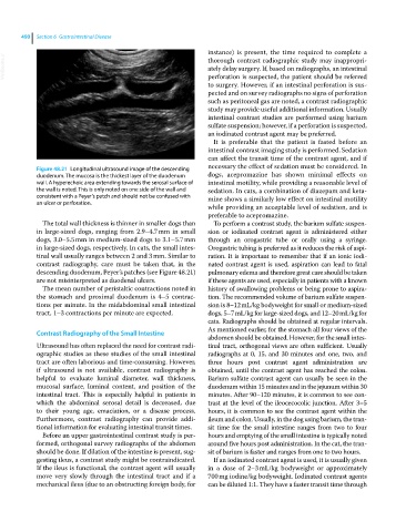
Figure 48.21 Longitudinal ultrasound image of the descending necessary the effect of sedation must be considered. In
duodenum. The mucosa is the thickest layer of the duodenum dogs, acepromazine has shown minimal effects on
wall. A hyperechoic area extending towards the serosal surface of intestinal motility, while providing a reasonable level of
the wall is noted. This is only noted on one side of the wall and sedation. In cats, a combination of diazepam and keta
consistent with a Peyer’s patch and should not be confused with mine shows a similarly low effect on intestinal motility
an ulcer or perforation.
while providing an acceptable level of sedation, and is
preferable to acepromazine.
The total wall thickness is thinner in smaller dogs than To perform a contrast study, the barium sulfate suspen
in large‐sized dogs, ranging from 2.9–4.7 mm in small sion or iodinated contrast agent is administered either
dogs, 3.0–5.5 mm in medium‐sized dogs to 3.1–5.7 mm through an orogastric tube or orally using a syringe.
in large‐sized dogs, respectively. In cats, the small intes Orogastric tubing is preferred as it reduces the risk of aspi
tinal wall usually ranges between 2 and 3 mm. Similar to ration. It is important to remember that if an ionic iodi
contrast radiography, care must be taken that, in the nated contrast agent is used, aspiration can lead to fatal
descending duodenum, Peyer’s patches (see Figure 48.21) pulmonary edema and therefore great care should be taken
are not misinterpreted as duodenal ulcers. if these agents are used, especially in patients with a known
The mean number of peristaltic contractions noted in history of swallowing problems or being prone to aspira
the stomach and proximal duodenum is 4–5 contrac tion. The recommended volume of barium sulfate suspen
tions per minute. In the midabdominal small intestinal sion is 8–12 mL/kg bodyweight for small or medium‐sized
tract, 1–3 contractions per minute are expected. dogs, 5–7 mL/kg for large‐sized dogs, and 12–20 mL/kg for
cats. Radiographs should be obtained at regular intervals.
As mentioned earlier, for the stomach all four views of the
Contrast Radiography of the Small Intestine
abdomen should be obtained. However, for the small intes
Ultrasound has often replaced the need for contrast radi tinal tract, orthogonal views are often sufficient. Usually
ographic studies as these studies of the small intestinal radiographs at 0, 15, and 30 minutes and one, two, and
tract are often laborious and time‐consuming. However, three hours post contrast agent administration are
if ultrasound is not available, contrast radiography is obtained, until the contrast agent has reached the colon.
helpful to evaluate luminal diameter, wall thickness, Barium sulfate contrast agent can usually be seen in the
mucosal surface, luminal content, and position of the duodenum within 15 minutes and in the jejunum within 30
intestinal tract. This is especially helpful in patients in minutes. After 90–120 minutes, it is common to see con
which the abdominal serosal detail is decreased, due trast at the level of the ileocecocolic junction. After 3–5
to their young age, emaciation, or a disease process. hours, it is common to see the contrast agent within the
Furthermore, contrast radiography can provide addi ileum and colon. Usually, in the dog using barium, the tran
tional information for evaluating intestinal transit times. sit time for the small intestine ranges from two to four
Before an upper gastrointestinal contrast study is per hours and emptying of the small intestine is typically noted
formed, orthogonal survey radiographs of the abdomen around five hours post administration. In the cat, the tran
should be done. If dilation of the intestine is present, sug sit of barium is faster and ranges from one to two hours.
gesting ileus, a contrast study might be contraindicated. If an iodinated contrast agent is used, it is usually given
If the ileus is functional, the contrast agent will usually in a dose of 2–3 mL/kg bodyweight or approximately
move very slowly through the intestinal tract and if a 700 mg iodine/kg bodyweight. Iodinated contrast agents
mechanical ileus (due to an obstructing foreign body, for can be diluted 1:1. They have a faster transit time through