
Page 519 - Clinical Small Animal Internal Medicine
P. 519
48 Gastrointestinal Imaging 487
Inflammatory Conditions of the Stomach
VetBooks.ir and Duodenum
Imaging Features of Gastritis
On survey radiographs, large rugae may be noted; this
may be most noticeable at the greater curvature of the
stomach. On a contrast radiographic study, large rugae
extending luminally and resulting in contrast‐sparing
areas are often present.
Sonographically, focal or circumferential wall thick
ening, loss of wall layering and, especially in chronic
disease, a mass effect can be noted. Regional lymph
node involvement may be present. Inflammatory
disease of the stomach may appear similar to neoplastic
or immune‐mediated disease or uremic gastropathy on
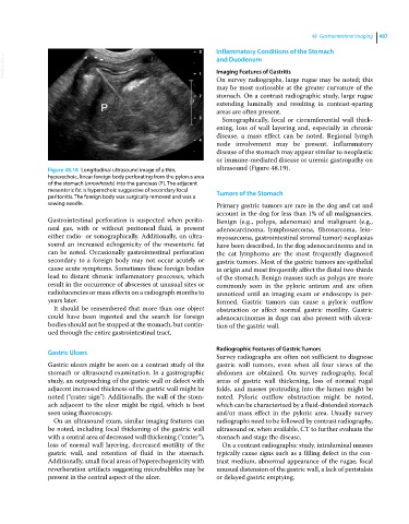
Figure 48.18 Longitudinal ultrasound image of a thin, ultrasound (Figure 48.19).
hyperechoic, linear foreign body perforating from the pylorus area
of the stomach (arrowheads) into the pancreas (P). The adjacent
mesenteric fat is hyperechoic suggestive of secondary focal Tumors of the Stomach
peritonitis. The foreign body was surgically removed and was a
sewing needle. Primary gastric tumors are rare in the dog and cat and
account in the dog for less than 1% of all malignancies.
Gastrointestinal perforation is suspected when perito Benign (e.g., polyps, adenomas) and malignant (e.g.,
neal gas, with or without peritoneal fluid, is present adenocarcinoma, lymphosarcoma, fibrosarcoma, leio
either radio‐ or sonographically. Additionally, on ultra myosarcoma, gastrointestinal stromal tumor) neoplasias
sound an increased echogenicity of the mesenteric fat have been described. In the dog adenocarcinoma and in
can be noted. Occasionally gastrointestinal perforation the cat lymphoma are the most frequently diagnosed
secondary to a foreign body may not occur acutely or gastric tumors. Most of the gastric tumors are epithelial
cause acute symptoms. Sometimes these foreign bodies in origin and most frequently affect the distal two‐thirds
lead to distant chronic inflammatory processes, which of the stomach. Benign masses such as polyps are more
result in the occurrence of abscesses at unusual sites or commonly seen in the pyloric antrum and are often
radiolucencies or mass effects on a radiograph months to unnoticed until an imaging exam or endoscopy is per
years later. formed. Gastric tumors can cause a pyloric outflow
It should be remembered that more than one object obstruction or affect normal gastric motility. Gastric
could have been ingested and the search for foreign adenocarcinomas in dogs can also present with ulcera
bodies should not be stopped at the stomach, but contin tion of the gastric wall.
ued through the entire gastrointestinal tract.
Radiographic Features of Gastric Tumors
Gastric Ulcers
Survey radiographs are often not sufficient to diagnose
Gastric ulcers might be seen on a contrast study of the gastric wall tumors, even when all four views of the
stomach or ultrasound examination. In a gastrographic abdomen are obtained. On survey radiography, focal
study, an outpouching of the gastric wall or defect with areas of gastric wall thickening, loss of normal rugal
adjacent increased thickness of the gastric wall might be folds, and masses protruding into the lumen might be
noted (“crater sign”). Additionally, the wall of the stom noted. Pyloric outflow obstruction might be noted,
ach adjacent to the ulcer might be rigid, which is best which can be characterized by a fluid‐distended stomach
seen using fluoroscopy. and/or mass effect in the pyloric area. Usually survey
On an ultrasound exam, similar imaging features can radiographs need to be followed by contrast radiography,
be noted, including focal thickening of the gastric wall ultrasound or, when available, CT to further evaluate the
with a central area of decreased wall thickening (“crater”), stomach and stage the disease.
loss of normal wall layering, decreased motility of the On a contrast radiographic study, intraluminal masses
gastric wall, and retention of fluid in the stomach. typically cause signs such as a filling defect in the con
Additionally, small focal areas of hyperechogenicity with trast medium, abnormal appearance of the rugae, focal
reverberation artifacts suggesting microbubbles may be unusual distension of the gastric wall, a lack of peristalsis
present in the central aspect of the ulcer. or delayed gastric emptying.