
Page 516 - Clinical Small Animal Internal Medicine
P. 516
484 Section 6 Gastrointestinal Disease
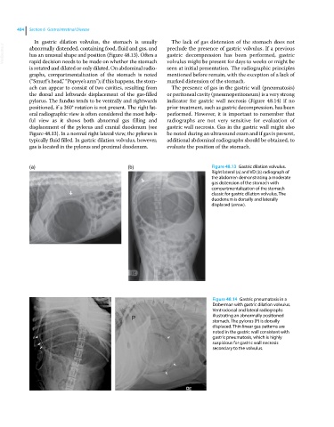
In gastric dilation volvulus, the stomach is usually The lack of gas distension of the stomach does not
VetBooks.ir abnormally distended, containing food, fluid and gas, and preclude the presence of gastric volvulus. If a previous
gastric decompression has been performed, gastric
has an unusual shape and position (Figure 48.13). Often a
rapid decision needs to be made on whether the stomach
seen at initial presentation. The radiographic principles
is rotated and dilated or only dilated. On abdominal radio volvulus might be present for days to weeks or might be
graphs, compartmentalization of the stomach is noted mentioned before remain, with the exception of a lack of
(“Smurf’s head,” “Popeye’s arm”); if this happens, the stom marked distension of the stomach.
ach can appear to consist of two cavities, resulting from The presence of gas in the gastric wall (pneumatosis)
the dorsal and leftwards displacement of the gas‐filled or peritoneal cavity (pneumoperitoneum) is a very strong
pylorus. The fundus tends to be ventrally and rightwards indicator for gastric wall necrosis (Figure 48.14) if no
positioned, if a 360° rotation is not present. The right lat prior treatment, such as gastric decompression, has been
eral radiographic view is often considered the most help performed. However, it is important to remember that
ful view as it shows both abnormal gas filling and radiographs are not very sensitive for evaluation of
displacement of the pylorus and cranial duodenum (see gastric wall necrosis. Gas in the gastric wall might also
Figure 48.13). In a normal right lateral view, the pylorus is be noted during an ultrasound exam and if gas is present,
typically fluid filled. In gastric dilation volvulus, however, additional abdominal radiographs should be obtained, to
gas is located in the pylorus and proximal duodenum. evaluate the position of the stomach.
(a) (b) Figure 48.13 Gastric dilation volvulus.
Right lateral (a) and VD (b) radiograph of
the abdomen demonstrating a moderate
gas distension of the stomach with
compartmentalization of the stomach
classic for gastric dilation volvulus. The
duodenum is dorsally and laterally
displaced (arrow).
Figure 48.14 Gastric pneumatosis in a
Doberman with gastric dilation volvulus.
Ventrodorsal and lateral radiographs
illustrating an abnormally positioned
stomach. The pylorus (P) is dorsally
displaced. Thin linear gas patterns are
noted in the gastric wall consistent with
gastric pneumatosis, which is highly
suspicious for gastric wall necrosis
secondary to the volvulus.