
Page 511 - Clinical Small Animal Internal Medicine
P. 511
48 Gastrointestinal Imaging 479
Figure 48.8 Neoplasia of the caudal
VetBooks.ir soft tissue attenuating mass is noted. On a
esophagus. In the caudal mediastinum, a
barium esophagram, ventral displacement
of the esophagus and an irregular contrast
pattern are noted outlining a soft tissue
attenuating structure. A small amount of
contrast agent is noted in the stomach.
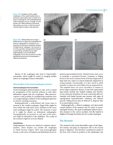
Figure 48.9 Sliding hiatal hernia (type 1 (a) (b)
hiatal hernia) in a dog, which presented for
thoracic radiographs to evaluate for the
presence of pulmonary metastatic disease.
(a) Right lateral radiograph. The stomach is
in normal position but part of the stomach
is summating cranially with the right
diaphragmatic crus. (b) Left lateral
radiograph. Part of the stomach is cranially
positioned (arrow) and the esophagus is
dilated (arrowheads).
Masses of the esophagus may lead to hypertrophic peritoneopericardial hernias. Hiatal hernias may occur
osteopathy, which might be noted on imaging studies as transient or persistent hernias. Transient or sliding
before an esophagus lesion is identified. hernia is the most common form of hernia diagnosed in
dogs and cats, where it is most frequently diagnosed in
young animals. Occasionally, it is also noted in adult
Abnormalities of the Gastroesophageal Junction animals, in whom it is more likely to be an acquired form.
Gastroesophageal Intussusception The acquired form can occur secondary to trauma or
Gastroesophageal intussusception is rare, and is caused severe upper respiratory disease. Commonly represented
by invagination of the stomach without or with other dog breeds include the English bulldog and SharPei.
abdominal organs into the esophagus. This intussus In cats, domestic shorthairs are more commonly affected.
ception is only possible if a predisposing condition such Animals with hiatal hernias may present with signs of
as a megaesophagus, dilation of the esophageal sphincter regurgitation, hypersalivation, vomiting, and delayed
or chronic vomiting is present. growth. Sliding hernia may be difficult to diagnose due
Radiographically, a well‐defined soft tissue mass is to its intermittent nature.
present in the caudal esophagus. Rugal folds might be Radiographically, a dilated esophagus and abnormally
seen within the soft tissue mass. Dilation of the more cranial position of the stomach may be noted. In parae
orad esophagus might be noted. The stomach or part sophageal hernias, the gastroesophageal sphincter/junc
of it might be absent on abdominal radiographs. tion remains in the correct position, but the fundus is
Sonographically, no stomach or only part of the stom displaced through the hiatus into the caudal esophagus.
ach might be identified in the abdomen. The cardia of
the stomach might be severely dilated.
The Stomach
Hiatal Hernia
Diaphragmatic hernias are relatively common abnor The stomach is the most distensible organ of the diges
malities. These hernias can involve the esophagus tive system, and plays an important role in the second
in hiatal hernias (Figure 48.9) and paraesophageal phase of digestion. The stomach is positioned caudal of
hernia, or the entry of stomach and abdominal viscera in the liver and is fixed in position at the diaphragm and