
Page 518 - Clinical Small Animal Internal Medicine
P. 518
486 Section 6 Gastrointestinal Disease
stomach might not always cause obstructive signs, but
VetBooks.ir might be toxic due to containing, for example, heavy
metals such as zinc. Therefore, in patients with intravas
cular hemolysis, abdominal radiographs may be indi
cated to evaluate for the presence of metallic foreign
objects. It is important to remember that aluminum is
relatively radiolucent, unlike a lot of other metals, and is
therefore difficult to detect on a survey radiograph.
Foreign bodies that are not mineral or metal attenu
ating can be challenging to detect. If foreign body
ingestion is suspected, multiple views of the abdomen
should be obtained. The gravity dependence of the fluid
and gas in the stomach may help to outline a foreign
body. If no foreign body is identified, contrast
radiography, ultrasound or CT might provide more
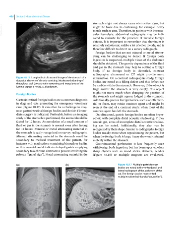
Figure 48.16 Longitudinal ultrasound image of the stomach of a information. On a contrast radiographic study, foreign
dog with a history of chronic vomiting. Moderate thickening of bodies are noted as a filling defect and this defect can
the pylorus wall (arrows) with narrowing and irregularity of the be mobile within the stomach. However, if the object is
luminal aspect is noted. D, duodenum.
large and/or the stomach is very empty, this object
might not move much when changing the position of
Foreign Bodies
the stomach and might appear lodged in the stomach.
Gastrointestinal foreign bodies are a common diagnosis Additionally, porous foreign bodies, such as cloth mate
in dogs and cats presenting for emergency veterinary rial or foam, may retain contrast agent and might be
care (Figure 48.17). It can often be a challenge to diag seen at the end of a contrast study, when most of the
nose gastrointestinal foreign bodies and decide if imme contrast agent has left the stomach.
diate surgery is indicated. Preferably, before an imaging On ultrasound, gastric foreign bodies are often hyper
study of the stomach is performed, the animal should be echoic with complete distal acoustic shadowing. If they
fasted for 12 hours. Accumulation of a small amount of contain gas, areas of incomplete distal acoustic shadow
fluid or gas in the stomach is normal even after fasting ing can be noted. Additionally, they also may be
for 12 hours. Mineral or metal attenuating material in recognized by their shape. Similar to radiography, foreign
the stomach is easily recognized on survey radiographs. bodies usually move when repositioning the patient, but
Mineral attenuating material in the stomach could be when the foreign body is large, it may show only minimal
secondary to medical treatment of the patient, for mobility within the stomach.
instance with medications containing bismuth or kaolin, Gastrointestinal perforation is less frequently seen
or this material could indicate delayed gastric emptying with foreign body ingestion, but has been reported when
secondary to a chronic obstructive process involving the sharp objects such as wood sticks, skewers, needles
pylorus (“gravel sign”). Metal attenuating material in the (Figure 48.18) or multiple magnets are swallowed.
(a) (b) Figure 48.17 Multiple gastric foreign
bodies are noted in the ventrodorsal and
lateral radiograph of the abdomen of the
cat. The foreign bodies represented
multiple rubber hair bands (“scrunchies”).