
Page 523 - Clinical Small Animal Internal Medicine
P. 523
48 Gastrointestinal Imaging 491
the intestinal tract, and reach the colon in approximately Inflammatory Disorders of the Small Intestine
VetBooks.ir half the time when compared with barium sulfate con Inflammatory bowel disease is a broad term encom
trast agents. As the iodinated contrast agents transit
passing all disease resulting in an inflammatory process
more rapidly through the gastrointestinal tract, radio
graphic views should be obtained more frequently, usu of the intestinal wall. On survey radiographs, usually no
abnormality is noted. It is important to remember that
ally every 10–30 minutes. As mentioned earlier, barium on survey radiographs, an increase in thickness of the
sulfate is inert and not metabolized by the gastrointesti small intestinal wall is difficult to impossible to assess
nal tract; however, iodinated contrast agents are absorbed as the presence of fluid and mucus within the intestinal
by the mucosa of the intestinal tract and therefore the tract can mimic intestinal wall thickening in loops of
luminal contrast rapidly declines over time, making diag intestine with normal wall thickness. On contrast radi
nosis of an intestinal lesion more difficult. ographic studies, a rapid transit of barium contrast
In the normal duodenum of the dog, small indenta
tions of the contrast agent (“pseudoulceration”) might be agent might be noted. Additionally, the mucosal surface
might be irregular and there might be a loss of normal
seen in the antimesenteric border of the wall. This is fimbriation of the mucosa. Enteritis may be generalized
normal, and results from the presence of Peyer’s patches. or only involve segments of the small intestinal tract.
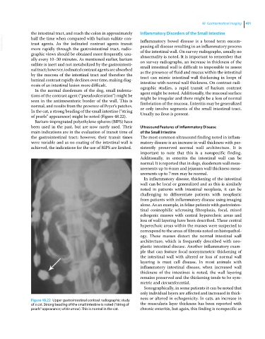
In the cat, a strong beading of the small intestine (“string Usually no ileus is present.
of pearls” appearance) might be noted (Figure 48.22).
Barium‐impregnated polyethylene spheres (BIPS) have
been used in the past, but are now rarely used. Their Ultrasound Features of Inflammatory Disease
main indications are in the evaluation of transit time of of the Small Intestine
the gastrointestinal tract; however, their transit times The most common ultrasound finding noted in inflam
were variable and as no coating of the intestinal wall is matory disease is an increase in wall thickness with per
achieved, the indications for the use of BIPS are limited. sistently preserved normal wall architecture. It is
important to note that this is a nonspecific finding.
Additionally, in enteritis the intestinal wall can be
normal. It is reported that in dogs, duodenum wall meas
urements up to 6 mm and jejunum wall thickness meas
urements up to 7 mm may be normal.
In inflammatory disease, thickening of the intestinal
wall can be local or generalized and as this is similarly
noted in patients with intestinal neoplasia, it can be
challenging to differentiate patients with neoplastic
from patients with inflammatory disease using imaging
alone. As an example, in feline patients with gastrointes
tinal eosinophilic sclerosing fibroplasia, focal, mixed
echogenic masses with central hyperechoic areas and
loss of wall layering have been described. These central
hyperechoic areas within the masses were suspected to
correspond to the areas of fibrosis noted on histopathol
ogy. These masses distort the normal intestinal wall
architecture, which is frequently described with neo
plastic intestinal disease. Another inflammatory exam
ple that can feature focal nonsymmetric thickening of
the intestinal wall with altered or loss of normal wall
layering is mast cell disease. In most animals with
inflammatory intestinal disease, when increased wall
thickness of the intestines is noted, the wall layering
remains preserved and the thickening tends to be sym
metric and circumferential.
Sonographically, in some patients it can be noted that
only individual layers are affected and increased in thick
ness or altered in echogenicity. In cats, an increase in
Figure 48.22 Upper gastrointestinal contrast radiographic study
of a cat. Strong beading of the small intestine is noted (“string of the muscularis layer thickness has been reported with
pearls” appearance; white arrow). This is normal in the cat. chronic enteritis, but again, this finding is nonspecific as