
Page 528 - Clinical Small Animal Internal Medicine
P. 528
496 Section 6 Gastrointestinal Disease
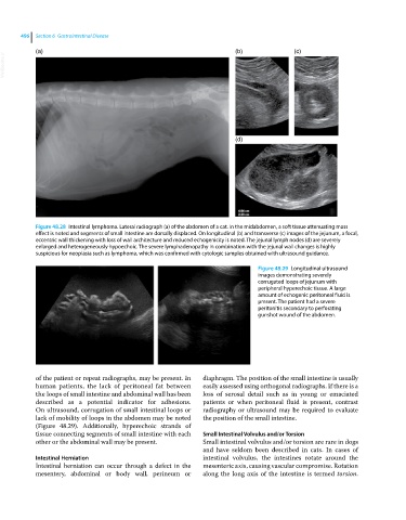
(a) (b) (c)
VetBooks.ir
(d)
Figure 48.28 Intestinal lymphoma. Lateral radiograph (a) of the abdomen of a cat. In the midabdomen, a soft tissue attenuating mass
effect is noted and segments of small intestine are dorsally displaced. On longitudinal (b) and transverse (c) images of the jejunum, a focal,
eccentric wall thickening with loss of wall architecture and reduced echogenicity is noted. The jejunal lymph nodes (d) are severely
enlarged and heterogeneously hypoechoic. The severe lymphadenopathy in combination with the jejunal wall changes is highly
suspicious for neoplasia such as lymphoma, which was confirmed with cytologic samples obtained with ultrasound guidance.
Figure 48.29 Longitudinal ultrasound
images demonstrating severely
corrugated loops of jejunum with
peripheral hyperechoic tissue. A large
amount of echogenic peritoneal fluid is
present. The patient had a severe
peritonitis secondary to perforating
gunshot wound of the abdomen.
of the patient or repeat radiographs, may be present. In diaphragm. The position of the small intestine is usually
human patients, the lack of peritoneal fat between easily assessed using orthogonal radiographs. If there is a
the loops of small intestine and abdominal wall has been loss of serosal detail such as in young or emaciated
described as a potential indicator for adhesions. patients or when peritoneal fluid is present, contrast
On ultrasound, corrugation of small intestinal loops or radiography or ultrasound may be required to evaluate
lack of mobility of loops in the abdomen may be noted the position of the small intestine.
(Figure 48.29). Additionally, hyperechoic strands of
tissue connecting segments of small intestine with each Small Intestinal Volvulus and/or Torsion
other or the abdominal wall may be present. Small intestinal volvulus and/or torsion are rare in dogs
and have seldom been described in cats. In cases of
Intestinal Herniation intestinal volvulus, the intestines rotate around the
Intestinal herniation can occur through a defect in the mesenteric axis, causing vascular compromise. Rotation
mesentery, abdominal or body wall, perineum or along the long axis of the intestine is termed torsion.